About this course:
The purpose of this module is to provide an overview of cervical precancer and cancer by outlining the core components of prevention, early detection, diagnosis, and management to inform APRN practice, facilitate prompt recognition and diagnosis, and improve clinical outcomes.
Course preview
The purpose of this module is to provide an overview of cervical precancer and cancer by outlining the core components of prevention, early detection, diagnosis, and management to inform APRN practice, facilitate prompt recognition and diagnosis, and improve clinical outcomes.
By the completion of this learning activity, the APRN should be able to:
- discuss the epidemiology of cervical cancer in the United States and risk factors for the development of the disease
- discuss the anatomy of the cervix and the pathophysiology leading to the development of cervical precancer and cancer
- summarize cervical cancer screening tests and early detection guidelines and review human papillomavirus (HPV) vaccine and immunization schedules
- identify the signs and symptoms of cervical cancer, components of the diagnostic workup, and clinical features as critical components of cervical cancer staging
- describe the management of cervical precancer and invasive cervical cancer, including an overview of treatment risks, side effects, and essential components of patient education
According to the American Cancer Society (ACS, 2020a), approximately 13,800 women in the United States will be diagnosed with invasive cervical cancer in 2020, and about 4,290 will die from the disease. Previously cited as one of the most common causes of cancer-related deaths, cervical cancer mortality rates have declined dramatically over the past 30 years with the advent and widespread utilization of the Papanicolaou (Pap) smear, followed by the addition of the HPV co-test. The recent arrival of the HPV vaccination has ignited a new era in cervical cancer prevention, demonstrating a substantial reduction in invasive cervical cancer since its inception. When diagnosed early and managed effectively, cervical cancer is highly treatable. APRNs practicing in primary care and women’s health settings must remain informed on evidence-based guidelines regarding the prevention, early detection, and management of precancerous lesions and invasive cervical cancers to facilitate an early diagnosis and reduce the morbidity and mortality associated with the disease (ACS, 2020a; Bruni et al., 2019).
Epidemiology
Cervical cancer is the fourth most common cancer among women, with 0.6% of women (1 in 159) diagnosed during their lifetime (Siegel et al., 2020). According to the World Health Organization (WHO, 2020), the annual age-adjusted incidence rate of cervical cancer is 7.6 per 100,000 people. Cervical cancer incidence is 30% higher in non-Hispanic Black (NHB) women than non-Hispanic White women (NHW; ACS, 2019). The states with the highest annual incidence and mortality rates include California (1,630 diagnoses/510 deaths), Texas (1,410/430), Florida (1,130/360), and New York (930/260), respectively (Cancer Statistics Center, 2020). The average age of diagnosis is 50, and the disease occurs most frequently in women aged 35 to 44 years. While 20% of cases occur in women over the age of 65, cervical cancer rarely develops in those who have maintained compliance with routine cervical cancer screenings (ACS, 2020a). Overall, cervical cancer ranks as the 12th leading cause of female cancer deaths in the United States; it is the second leading cause of cancer deaths in women aged 20 to 39 years (Siegel et al., 2020) and the third leading cause of cancer deaths in women aged 15 to 44 years (Bruni et al., 2019). The 5-year survival rate of localized cervical cancer (i.e., cancer that has not spread outside of the cervix or uterus) is 92%, declining to 17% for those with distant metastases (i.e., cancer spreading to organs or body parts outside of the cervix or uterus). NHB women are more likely to be diagnosed at advanced stages of the disease, reducing their 5-year survival rate to 56% compared to 68% in NHW women (ACS, 2019; 2020b). While survival has improved since the 1970s for most cancer types, it has remained relatively stagnant for cervical cancers; this is attributed to the lack of significant treatment advances for patients with recurrent and metastatic disease. Furthermore, nearly 90% of women who die from cervical cancer have inadequate access to prevention, screening, and treatment (WHO, 2020).
Risk Factors
Risk factors for cervical cancer include HPV infection, tobacco use, early onset of sexual activity, multiple sexual partners, and HIV infection. Predominantly transmitted through sexual contact, HPV is the chief risk factor for cervical cancer, leading to nearly 99% of diagnoses. Tobacco use is the only nonsexual behavior associated with cervical dysplasia (abnormal growth of cells on the cervix’s surface) and cancer, serving as a significant cofactor for the development of HPV. Women who smoke are at least twice as likely as non-smokers to be diagnosed with cervical cancer. Research has demonstrated that among women with HPV infections, those who smoke have a significantly higher viral load on the cervix, heightening the risk of cervical cancer development (Fang et al., 2018; Truth Initiative, 2019). Early sexual activity and multiple sexual partners (especially those who have had numerous partners themselves) increase the risk of HPV exposure. People who are immunocompromised—such as those with HIV, transplant recipients, cancer patients, and those receiving immunosuppressive medications—are at higher risk for acquiring an HPV infection (ACS, 2020a).
Pathophysiology
The cervix is the fibromuscular lower portion of the uterus (the hollow, pear-shaped organ that carries a fetus) that connects the uterine cavity to the vagina, as demonstrated in Figure 1. It measures 4 cm in length and 3 cm in diameter in an adult woman of childbearing age and undergoes progressive involution following menopause. The cervix is substantially larger in parous women (i.e., those who have given birth) than nulliparous women (i.e., those who have never carried a pregnancy to term; McCance & Heuther, 2019; Mehta & Sachdeva, 2017).
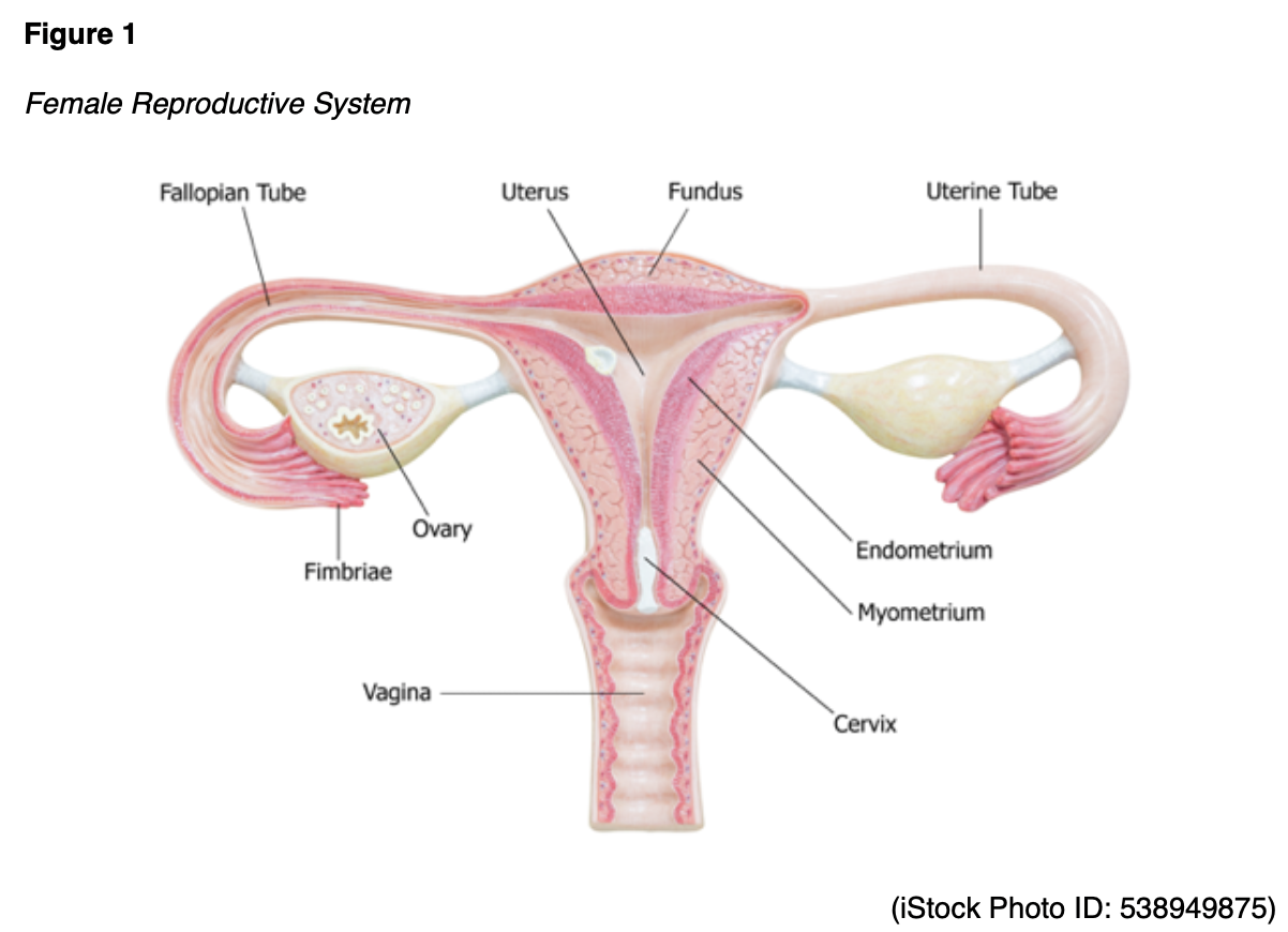
Two sections comprise the cervix: the ectocervix and the endocervix. The ectocervix extends into the vagina and is lined with stratified squamous non-keratinized epithelium. The squamous epithelium involves four layers: basal, parabasal, intermediate, and superficial. As demonstrated in Figure 2, the cervix opens into the vagina through the external os (external orifice), which is the readily visible portion of the cervix. The external os marks the transition point to the endocervix (endocervical canal), which is the inner part of the cervix lined by mucus-secreting columnar epithelium or glandular cells. The endocervix is rarely visible on an internal pelvic examination. It forms a passageway between the external and internal os (internal orifice or isthmus). The internal os is a narrow opening that marks the transition from the endocervix to the endometrium, which is the lining of the uterine cavity. The cervix enables the passage of sperm into the uterine cavity through dilation of the external and internal os. The cervix maintains a sterile environment by frequent shedding of the endometrium, thick cervical mucus production, and a narrow external os to protect against bacterial invasion (McCance & Heuther, 2019; Mehta & Sachdeva, 2017).

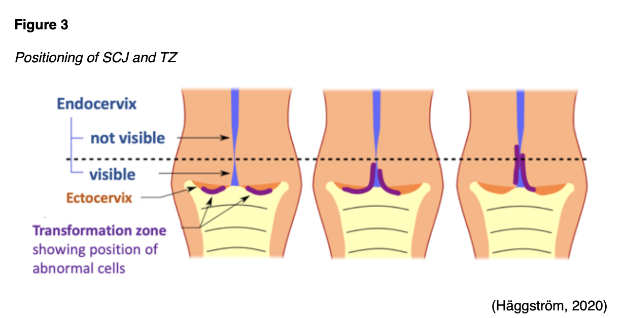
The area between the endocervix and the ectocervix is called the squamocolumnar jun
...purchase below to continue the course
HPV is a ubiquitous group of tiny viruses that carry deoxyribonucleic acid (DNA) that can adapt to their host and evade the immune system. An estimated 12,000 new cases of HPV-associated cervical cancers are diagnosed in the United States each year (Centers for Disease Control and Prevention [CDC], 2020b). A growing body of evidence links HPV to other anogenital cancers (anal, vulvar, vaginal, penile), as well as head and neck cancers (Bruni et al., 2019). Of the 200 identified strains of HPV, approximately 40 have a particular affinity for the genital region. While most of these are low-risk and non-oncogenic (wart-causing), at least 13 are high-risk and oncogenic (cancer-causing), leading to cervical cancer (CDC, 2020a; Wang et al., 2018).
The most common drivers of cervical carcinogenesis are the high-risk subtypes HPV-16 and HPV-18, which are responsible for at least 70% of cervical cancers (Bruni et al., 2019; WHO, 2019). The majority of sexually active males and females will be infected with HPV at some point during their lives, but most of these infections will remain clinically silent. The immune system often mediates the spontaneous clearance of HPV from the cervix within 2 years of acquiring the infection. However, about 10% of women with HPV will develop a persistent infection (Mello & Sundstrom, 2020). Chronic infections with high-risk HPV subtypes can lead to cervical precancers or cervical intraepithelial neoplasia (CIN). If it is left untreated, CIN can invade local tissues and progress to invasive cancer (CDC, 2020a; Hahn & Spach, 2018).
The Role of HPV in Cervical Carcinogenesis
HPV leads to CIN and invasive cancer through a series of pathologic processes in which oncogenes and tumor suppressor genes serve fundamental roles. An oncogene is a mutation associated with cancer development; it can be inherited or caused by exposure to carcinogenic substances in the environment, such as ultraviolet (UV) radiation and free radicals. In their normal and non-mutated state, oncogenes are called proto-oncogenes, regulating healthy cell growth and division. When a proto-oncogene mutates into an oncogene, it becomes permanently activated (turned on), fueling unregulated cell growth and resultant cancer cells. Tumor suppressor genes are healthy genes that slow cell division, repair DNA errors, and induce apoptosis (programmed cell death). Under physiologic conditions, tumor suppressor genes regulate healthy cellular growth and division and prevent cells with mutated or damaged DNA from replicating. When these tumor suppressor genes are inactivated (turned off), these processes become unregulated, thereby fueling cancer growth (CancerQuest, 2020).
The HPV genome comprises three regions: the early gene-coding region (E), the late gene-coding region (L), and the long control region (LCR). The focal point for HPV-carcinogenesis occurs within the early genes (E1, E2, E4, E5, E6, and E7). The early genes encrypt for viral replication. Oncogenic HPV subtypes encode two primary viral oncoproteins (E6 and E7), which govern the initial changes in cervical epithelial tissue. Infection with HPV arises at the stratified squamous epithelial cells’ basal cell layer and stimulates cellular proliferation. Overexpression of E6 and E7 facilitates the malignant transformation of high-risk subtypes of HPV by disrupting DNA repair mechanisms and apoptosis, leading to rapid and unregulated cell proliferation. The growth of HPV-positive cancer cells relies on the sustained expression of E6 and E7, as the virus is incapable of replicating on its own; it depends on the cellular division and stratification of the epithelium to replicate and produce new viruses. E6 and E7 work in concert to inactivate their respective substrates, p53 and retinoblastoma (pRb), which are two major tumor suppressors. Inactivation of these substrates leads to ongoing cellular proliferation. Unregulated overexpression of the E6 and E7 oncoproteins leads to changes in the cellular pathways and genes involved in DNA repair, growth factors, and angiogenesis (the development of new blood vessels). These changes allow the virus to amplify its genome, complete its life cycle, and produce new virions. Without proper treatment, HPV-infected cells will progress to invasive cervical cancer. The malignant transformation process and the role of HPV are demonstrated in Figure 4 (Hahn & Spach, 2018; Pal & Kundu, 2020).

Pap Smear and HPV Testing
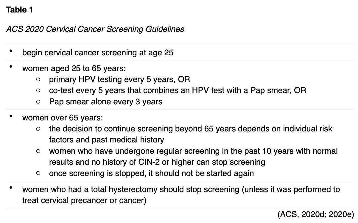
Since HPV-induced CIN does not cause symptoms, cellular changes are identified on screening Pap smear and/or HPV testing. A Pap smear is a noninvasive test to evaluate for cervical abnormalities, precancerous lesions, and invasive cancer. It can also diagnose benign conditions such as infection and inflammation, but it does not diagnose HPV itself. HPV can be detected about a year after infection in the cervix, and HPV testing is a critical component of cervical cancer screening. In the United States, cervical cancer screening guidelines support primary HPV testing alone or HPV co-testing. Primary HPV testing is a relatively novel screening modality performed alone, whereas HPV co-testing involves Pap and HPV tests that are performed together. The ACS (2020e) recommends the primary HPV test for cervical cancer screening for women aged 25 to 65. In contrast, the American Society for Colposcopy and Cervical Pathology (ASCCP) supports the use of either test, endorsing any type of HPV–based testing as the basis for risk estimation (Perkins et al., 2019). Table 1 provides a basic overview of the 2020 ACS cervical cancer screening guidelines.
The U.S. Food & Drug Administration (FDA) has approved specific primary HPV tests, including target- and signal-amplification assays of HPV DNA, as well as HPV mRNA. Most FDA-approved primary HPV assays have demonstrated similar performance metrics regarding efficacy across clinical trials; however, they are not yet widely available or adopted across practice settings. Therefore, HPV co-testing remains the most common screening test among women aged 30 to 65 years in the United States (Arbyn et al., 2015; Perkins et al., 2020). Regardless of the modality, all HPV tests evaluate the cells’ internal environment explicitly for signs of infection with high-risk viral subtypes. Viral nucleic acid (DNA or mRNA) must be detected within the cervical cells to form a definitive diagnosis. As shown in Figure 5, to perform a Pap smear, the clinician positions a vaginal speculum to visualize the cervix and uses a small, cone-shaped brush and plastic spatula to scrape the cervix and collect cells. Next, the brush and spatula are rinsed in a liquid-filled vial and sent to the laboratory for processing. Most women tolerate the test without any complication or pain. Slight vaginal spotting may occur immediately following the Pap smear, which resolves in under 24 hours (National Cancer Institute [NCI], 2019b).

Pap Smear Results
The results of the Pap smear denote whether the cervical cells are normal or abnormal, but some samples may return as unsatisfactory (see below). All Pap smears must include cells within the SCJ.
Normal Results
Normal (negative) results indicate no abnormal cells were identified, and patients do not require further workup or management. Patients are usually advised to undergo routine screening in 3 to 5 years, according to evidence-based cervical cancer screening guidelines.
Unsatisfactory Results
Unsatisfactory Pap smear results indicate that there were insufficient cells to evaluate the specimen definitively, or the cells may have been obscured by blood or mucus. Patients with unsatisfactory results typically require repeat testing.
Abnormal Results
Abnormal (positive) results indicate abnormal cellular changes in the sample and any grade of dysplasia warrants further workup. A pathologist classifies the severity of the changes based on their risk of progressing to invasive cancer. There are several types of abnormal Pap results; the most common include the following:
- Atypical squamous cells of undetermined significance (ASCUS)
- ASCUS is the most common abnormal Pap result and confirms the presence of abnormal cells. ASCUS is an inconclusive finding that does not determine the origin of the abnormal cells or whether the changes are secondary to an HPV infection. Several benign causes of atypical-appearing cells include yeast infection, irritation, polyps, cysts, and hormonal changes during pregnancy and menopause. The management of ASCUS typically includes HPV co-testing or a repeat Pap smear in 1 year.
- Atypical glandular cells (AGC)
- AGC signifies that some glandular cells do not appear normal.
- Squamous intraepithelial lesion (SIL)
- SIL refers to dysplastic changes in the cervix and is categorized into low-grade (LSIL) or high-grade (HSIL). LSIL denotes early cellular changes (mild dysplasia), meaning cells have only a few abnormal characteristics and still resemble healthy cells. HSIL denotes moderate or severe dysplasia, in which cells appear highly abnormal under the microscope but remain confined to the cervix’s surface. While HSIL does not indicate invasion into deeper parts of the cervix, it can develop into cervical cancer if it is not treated.
- Carcinoma in situ (CIS)
- CIS denotes early-stage noninvasive cancer, in which the cancerous cells arise from and are limited to the squamous epithelium. CIS has not penetrated more deeply into the tissues.
- Adenocarcinoma in situ (AIS)
- AIS indicates a noninvasive lesion of the glandular tissue and is much less common than CIS. AIS has not penetrated more deeply into the tissues.
- Invasive cervical cancer
- Less commonly, Pap smears can detect malignant cells (squamous or adenocarcinoma). However, invasive cervical cancer is rarely identified on a Pap smear in women who have undergone screening at regular intervals, as cervical abnormalities typically manifest as precancerous lesions (ACS, 2019; NCI, 2019b; Mello & Sundstrom, 2020; Perkins et al., 2020).
HPV Test Results
HPV test results are more simple than Pap smear results, as the goal is to detect high-risk HPV types. An HPV test result is either positive (high-risk HPV was found) or negative (high-risk HPV was not found; Arbyn et al., 2015; Perkins et al., 2020).
Follow-Up of Abnormal Results
Colposcopy
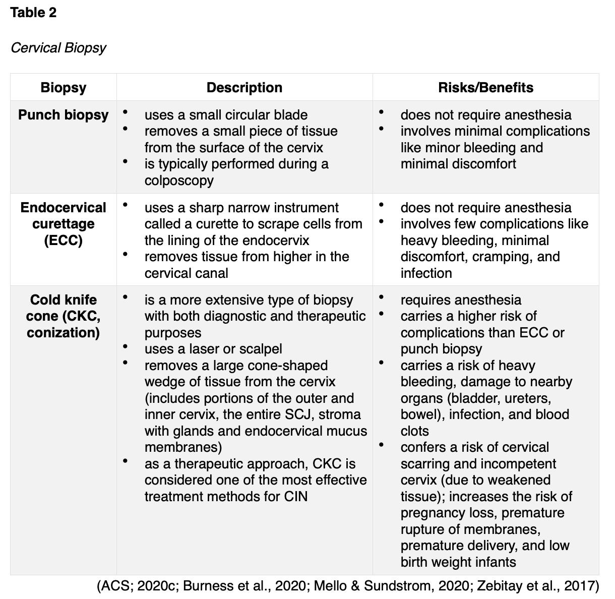
As demonstrated in Figure 6, colposcopy is a noninvasive follow-up procedure performed after abnormalities are detected on a Pap smear or from HPV testing. A colposcopy diagnoses precancerous changes and benign conditions, such as benign polyps, cervicitis (inflammation of the cervix), or genital warts. During the procedure, a weak acetic acid solution (diluted vinegar) is applied to the cervix. The vinegar turns abnormal areas white, allowing for direct visualization of cervical changes under a stereomicroscope. A biopsy of the abnormal area is performed, as tissue sampling and histological analysis are the only way to diagnose precancer or cancer definitively. There are three major types of cervical biopsies, as outlined in Table 2. A colposcopy is a safe procedure that carries minimal risks. The potential harms may include discomfort, psychological distress, infection, and bleeding (ACS, 2020c; Burness et al., 2020; Khan et al., 2017).

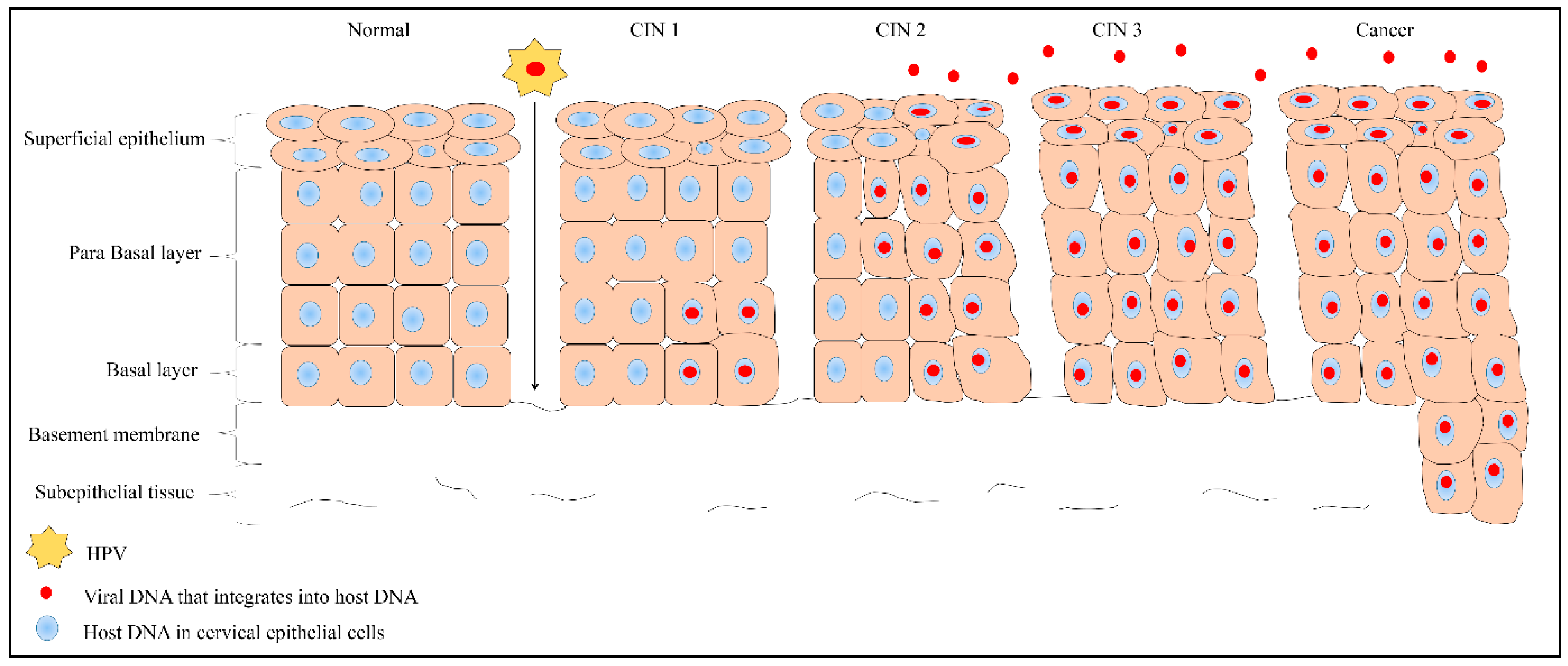
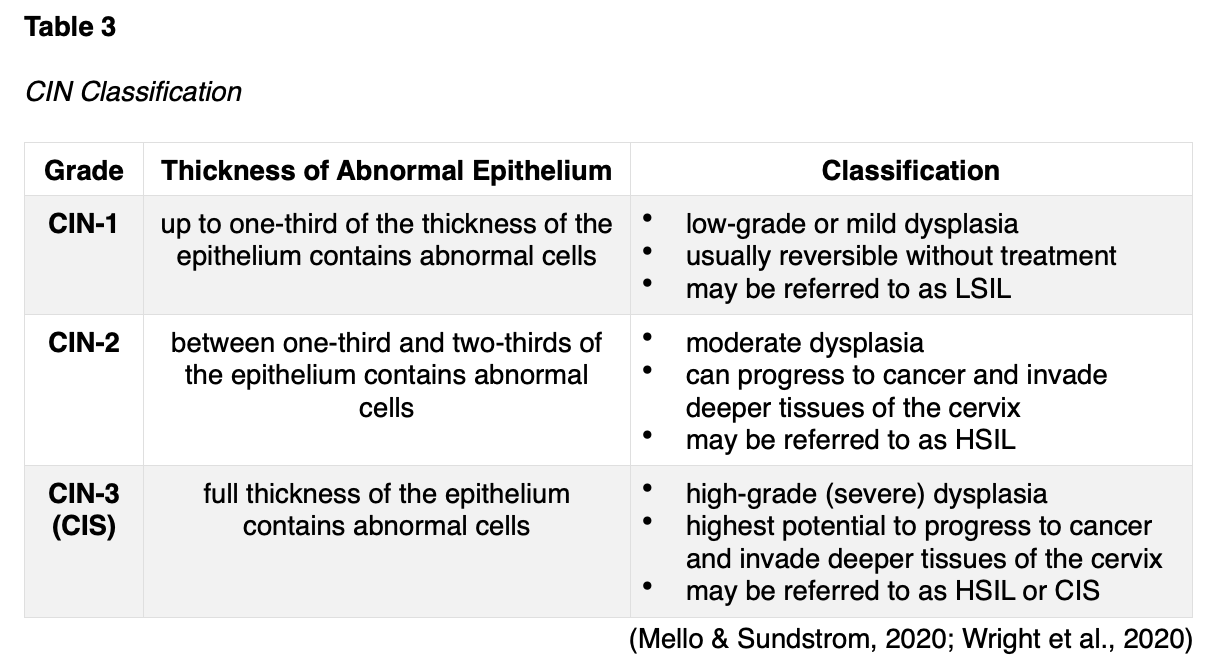
CIN is a histological diagnosis determined from a biopsy specimen. CIN is a noninvasive, precancerous condition in which the abnormal cells are confined to the epithelium and relates directly to the integration of an HPV infection. It denotes the extent of dysplasia within the layers of the tissue and is classified into three grades based on how deeply the abnormal cells invade into the skin. As the CIN grade advances, the risk for progression to invasive cancer heightens, and natural regression is uncommon. The classification of CIN is further delineated in Table 3. Figure 7 depicts the process by which early changes (CIN-1) progressively affect the deeper layers of the epithelium, leading to full-thickness involvement in its most severe form (CIN-3/CIS; Mello & Sundstrom, 2020).
Figure 7
Progression of CIN to Cancer
CIN Management and Treatment
CIN treatment aims to eradicate the precancerous lesions to prevent them from progressing to invasive cancer. Treatment selection depends on various factors such as the extent and severity of the CIN, the patient’s age, their medical history, cost-effectiveness, availability, and the potential risks and benefits of each method. Procedures to treat CIN can affect a woman’s future childbearing potential, so shared decision-making is crucial to ensure patients are fully informed of the potential risks before deciding on a therapy option. Most low-grade CINs resolve without intervention; only about 1% of cases will progress to cervical cancer. According to the ASCCP, most patients with CIN-1 can be adequately managed with close observation using Pap smears, HPV testing, and colposcopies as indicated. In contrast, patients with high-grade CIN typically require intervention. Treatment options include ablative and excisional techniques. Ablative techniques (e.g., cryotherapy) destroy abnormal tissues by freezing them, whereas excisional methods (e.g., loop electrosurgical excision procedure [LEEP] and CKC, described in Table 1) remove the abnormal tissue surgically. The WHO and the ASCCP strongly recommend excisional treatment over cryotherapy in all settings where a LEEP procedure is available and accessible. LEEP and CKC are the two most common and effective excisional procedures for CIN-2, CIN-3, and CIS (Perkins et al., 2020; Wright et al., 2020).
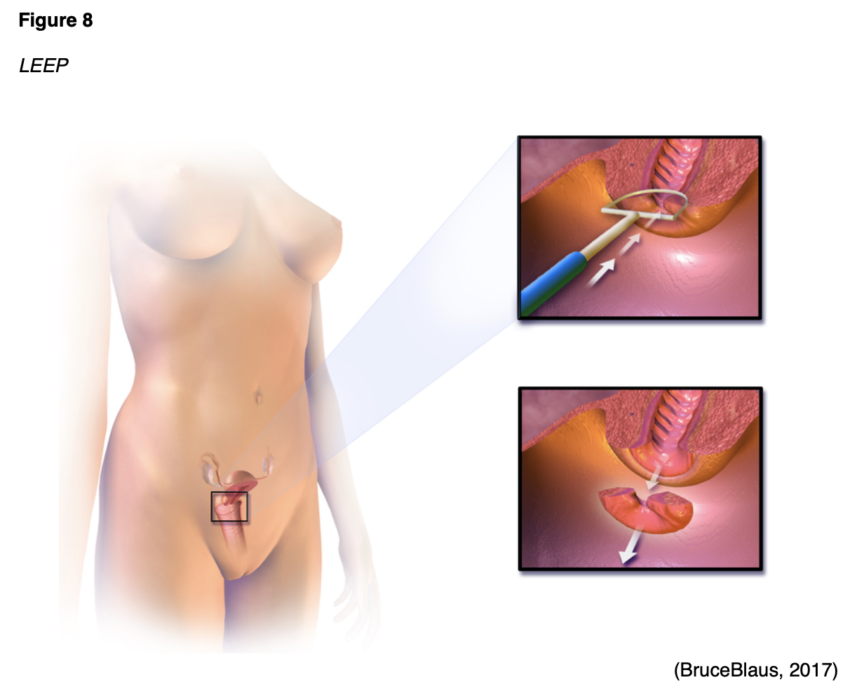
Loop Electrosurgical Excision Procedure (LEEP)
LEEP is a widely utilized treatment for high-grade cervical dysplasia following colposcopy. As shown in Figure 8, a thin wire loop that functions as a scalpel is used to excise abnormal cells from the cervix. An electric current is passed through the loop to remove a thin layer of the cervix. Patients may feel mild discomfort during the procedure, so local anesthesia is typically used for pain control. The procedure is usually performed in the outpatient setting and only takes about 15 minutes. At the end of the procedure, a reddish-brown paste called ferric subsulfate (Monsel’s solution) may be applied to the cervix to control any bleeding; alternatively, the blood vessels may be cauterized (heated with a device to control bleeding). If ferric subsulfate (Monsel’s solution) is used, a harmless brownish vaginal discharge may occur for up to 48 hours after the procedure. The most common side effects include mild discomfort and slight bleeding within the first 2 to 3 weeks after the procedure. To allow the cervix to heal fully, patients should avoid placing anything inside the vagina (including tampons and douching) and abstain from sexual intercourse for 4 to 6 weeks. Patients may shower as usual but should avoid baths. A LEEP is associated with a small increased risk for future pregnancy complications, including premature birth and low birth weight. In rare cases, the cervix may narrow after the procedure, causing menstrual irregularities (The American College of Obstetricians and Gynecologists, 2020; Perkins et al., 2020).
Cryotherapy
Cryotherapy, which is also referred to as cryosurgery or laser ablation, is demonstrated in Figure 9. It involves applying a cooled metal disc (cryoprobe) to the cervix to freeze the precancerous cells. The cryoprobe is cooled using compressed carbon dioxide (CO2) or nitrous oxide (NO) gas. Cryotherapy is a simple, fast outpatient procedure that is performed without anesthesia. It typically takes about 15 minutes and may cause mild discomfort. Following cryotherapy, the frozen area regenerates with normal epithelium in about a month, and some patients can experience watery vaginal discharge during this time. Patients should avoid sexual intercourse or placing anything in the vagina during the healing process for 4 to 6 weeks (Mello & Sundstrom, 2020; Perkins et al., 2020).
According to the ASCCP guidelines, cryotherapy should not be used in any of the following clinical circumstances:
- for lesions that extend into the canal,
- for lesions covering more than 75% of the surface area of the ectocervix,
- if the SCJ or upper limit of any lesion is not fully visualized,
- if the CIN cannot be graded or is CIN-2 or higher,
- after prior treatment for CIN-2 or higher, or
- in the setting of inadequate biopsies of the cervix to confirm a histologic diagnosis (Perkins et al., 2020, p. 118).

Surveillance Following CIN Treatment
The ASCCP supports continued surveillance with HPV testing or co-testing at 3-year intervals for at least 25 years after treatment for HSIL, CIN-2, CIN-3, or CIS. Extending surveillance beyond 25 years is acceptable if the patient’s life expectancy and screening capabilities are not compromised by concomitant health conditions (Perkins et al., 2019).
AIS Management
Although AIS frequently coexists with CIS, it is much less common and requires heightened management. The incidence of AIS has increased in recent decades among women aged 30 to 40. The average interval between the diagnosis of AIS and progression to early invasive cancer is about 5 years. While AIS may be detected on Pap smear, a definitive diagnosis requires tissue sampling via biopsy. AIS typically develops in the TZ and extends proximally to the endocervical canal. When diagnosed with a cervical biopsy, about 15% of patients have an invasive component (Teoh et al., 2020). The standard treatment for AIS is hysterectomy (removal of the uterus) unless future fertility is desired. If fertility is desired, excisional procedures (e.g., LEEP or CKC) are acceptable if the patient is willing to adhere to the following heightened surveillance recommendations: HPV co-testing and endocervical sampling every 6 months for at least 3 years, then annually for at least 2 years (or until hysterectomy). Once childbearing is complete, a hysterectomy should be performed (Perkins et al., 2020).
Cervical Cancer Subtypes
Cervical cancer ensues when precancerous cells invade the basement membrane. It occurs in two major subtypes: squamous cell carcinoma (SCC) and adenocarcinoma. The majority of cervical cancers (>80%) are SCC, originate in the squamous epithelium in the TZ, and closely correlate with chronic HPV infection as described earlier. Cervical adenocarcinoma begins in the glandular cells that line the upper portion of the cervix and cause up to 20% of cervical cancers. Adenocarcinomas originate in the TZ or the glandular epithelium above the TZ and are distinct from SCC, as they are not uniformly caused by infection with HPV (Bruni et al., 2019; Hodgson & Park, 2019). Neuroendocrine carcinoma of the cervix (NECC) is a third, rare, and more aggressive type that accounts for only 1.5% of all cervical cancers. There are four subtypes of NECC tumors: small cell neuroendocrine carcinoma (SCNEC), large cell neuroendocrine carcinoma (LCNEC), typical carcinoid tumor, and atypical carcinoid tumor. Of these, SCNEC is the most common and aggressive type and carries the poorest prognosis. In contrast, carcinoid tumors typically have a more indolent disease trajectory and a more favorable prognosis. The role of HPV in mediating the development of NECC tumors is not clearly understood. Unlike SCC and adenocarcinoma, NECC is not universally preceded by preinvasive disease. Therefore, early detection via Pap smear screening is less common. The management of NECC is more complex and less precise due to the rarity of the condition but typically includes multiple chemotherapy agents (Tempfer et al., 2018).
Signs and Symptoms
Women with early-stage cervical cancers typically do not have symptoms. With cervical cancer, symptoms do not develop until the cancer advances, grows larger, and invades nearby tissue. The most common symptoms include the following:
- abnormal vaginal bleeding, such as spotting between periods, bleeding after intercourse, menstrual periods that are longer or heavier than usual, and postmenopausal bleeding;
- dyspareunia (pain during or immediately following intercourse) or pain in the pelvis not related to intercourse;
- abnormal vaginal discharge;
- swelling of the legs due to impaired lymphatics caused by tumor compression;
- urinary symptoms such as frequency, dysuria, inability to void, or hematuria; and
- constipation (ACS, 2020a).
Diagnostic Workup
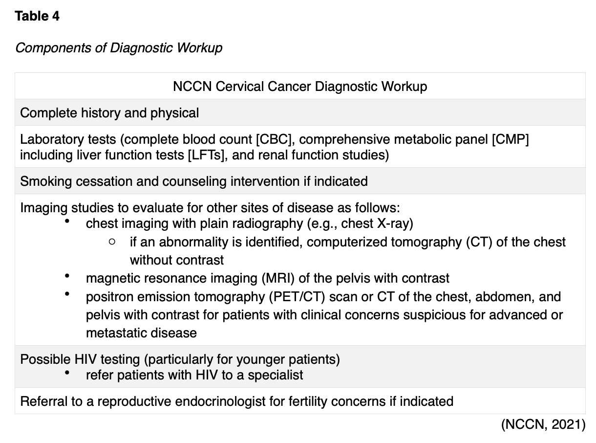
The diagnosis and management of cervical cancer are multifactorial and involve the use of combined modalities. Most institutions accredited by the NCI rely on evidence-based guidelines from the National Comprehensive Cancer Network (NCCN) to outline cancer diagnosis, staging, and treatment planning. The NCCN is an alliance of leading cancer centers and world-renowned experts devoted to cancer care, research, and education. Through rigorous clinical trial research, data compiled across institutions, and annual expert panel reviews, the NCCN provides evidence-based treatment guidelines for cancer according to subtype, pathology, genetics, staging, inheritance patterns, and other specific features. The guidelines are widely utilized in cancer care and guide medical decision making throughout the patient’s disease trajectory (NCCN, 2021). Once cervical cancer is diagnosed through biopsy and histologic analysis, the NCCN (2021) guidelines endorse various components of further diagnostic workup, which are outlined in Table 4.
Principles of Pathologic Review
The biopsy sample undergoes a series of tests to determine the cancer’s pathologic features, evaluate its behavior, and select the best treatment options. Tumor grade is a measurement of how different the cancer cells look from healthy cells under the microscope. The grading determines the likelihood of cancer growth and spread and is defined as follows:
- Grade 1 is well-differentiated (appears similar to healthy cells) and the least aggressive.
- Grade 2 is moderately differentiated, appears less like healthy cells, and is an intermediate grade (more aggressive than grade 1).
- Grade 3 is poorly differentiated or undifferentiated (does not resemble healthy cells at all), is high-grade and most aggressive, and tends to grow and spread more quickly (ACS, 2020a; Yarbro et al., 2018).
Gene and Molecular Biomarker Analyses
Tumor protein p53 (TP53)
Under physiologic conditions, TP53 functions as a tumor suppressor gene, regulating cellular growth and division by preventing cells from growing and dividing uncontrollably. TP53 keeps cells with mutated or damaged DNA from dividing, thereby preventing the development of tumors. Mutations in the TP53 impairs its ability to control cell proliferation, as it cannot trigger apoptosis in cells with mutated or damaged DNA. Consequently, DNA damage accumulates in cells, and the cells continue to divide in an uncontrolled way, leading to tumor growth. In cervical cancer, TP53 is inactivated by the HPV oncoprotein E6, thereby driving carcinogenesis (Nakamura et al., 2019; U.S. National Library of Medicine, 2020).
p16 Immunohistochemistry
P16 is a tumor suppressor protein that performs critical functions in regulating the normal cell cycle. Nearly all HPV-positive cervical precancers and cancers are accompanied by p16 overexpression. P16 interacts with HPV, inducing cell cycle dysfunction, interfering with apoptosis, facilitating cell invasion, and inducing angiogenesis. P16 overexpression paradoxically intensifies with increasing aggressiveness of the cancer. As highlighted throughout the literature, p16-negative tumors are associated with poorer outcomes and reduced survival compared to their p16-positive counterparts. Despite the widespread prevalence of p16 overexpression in cervical cancer, its clinical value is unclear. A lack of consensus or guidelines persists regarding the use of p16 overexpression in guiding treatment (Nicolas et al., 2020).
Mismatch Repair (MMR)/Microsatellite Instability (MSI)
Advancements in modern technology have led to novel treatment strategies utilizing the immune system to attack cancer. Specialized tests called microsatellite instability-high (MSI-H) testing and mismatch repair deficiency (dMMR) determine whether cancer is susceptible to the antitumor efforts of immune-based therapies. The presence of MSI-H or dMMR act as inhibitors to the anti-programmed cell death protein 1 (PD-1)/programmed cell death ligand 1 (PD-L1) pathway, which serves essential functions in regulating the immune system. The NCCN recommends that all patients with recurrent, progressive, or metastatic cervical cancer should undergo testing for MSI-H or dMMR. MMR deficiency can be reported as MSI-H or dMMR, but they share the same meaning (NCCN, 2021).
Cancer Staging
Worldwide, the staging system of the International Federation of Gynecology and Obstetrics (FIGO) guides cervical cancer staging. The guidelines were last updated in 2018 and are integrated within the NCCN guidelines. The four stages of cervical cancer are described in Table 5 and displayed in Figure 10. Since many cases require specialized surgical care, all patients with suspected cervical cancers should be promptly referred to a gynecologic oncologist (ACS, 2020b; Bhatla et al., 2018; NCCN, 2021).


Cervical Cancer Treatment
The most optimal cervical cancer treatment depends on various factors, such as the pathologic features, cancer stage, plans for fertility, patient preference, age, and medical history. Treatment is often multimodal, with several therapies combined and administered simultaneously (concurrently) or sequentially. This section will provide a synopsis of the most common evidence-based treatment strategies (ACS, 2020a; NCCN, 2021).
Surgery
Surgical intervention for cervical cancer can be fertility-sparing or non-fertility-sparing. Most early-stage cervical cancers (stage IA1) can be effectively managed with LEEP or CKC, both of which are fertility-sparing treatments. In patients with stage IA2 or stage IB1, cancers may be managed with a radical trachelectomy with lymphadenectomy. A radical trachelectomy, also called a cervicectomy, involves the surgical removal of the cervix, upper vagina, and supporting ligaments, but the uterine corpus is preserved (see Figure 11). The surrounding pelvic lymph nodes are also removed to evaluate for cancer spread. With regard to its impact on future pregnancies, a radical trachelectomy is associated with a 10% likelihood of second-trimester loss, but more than 70% of women carry gestation to 37 weeks or more (NCCN, 2021).

When fertility preservation is not desired or irrelevant, early-stage cervical cancers are managed with radical hysterectomy. The standard of care for stage IA2, IB1, IB2, IB3, and IIA1 cervical cancers is a radical hysterectomy with bilateral pelvic lymphadenectomy or pelvic lymph node dissection (PLND). A radical hysterectomy (see Figure 12) involves removing the uterus, cervix, a portion of the vagina, and the parametrium (the connective tissue that surrounds the cervix). PLND refers to the removal of the lymph nodes from the pelvis; the ovaries and fallopian tubes remain intact (NCCN, 2021).

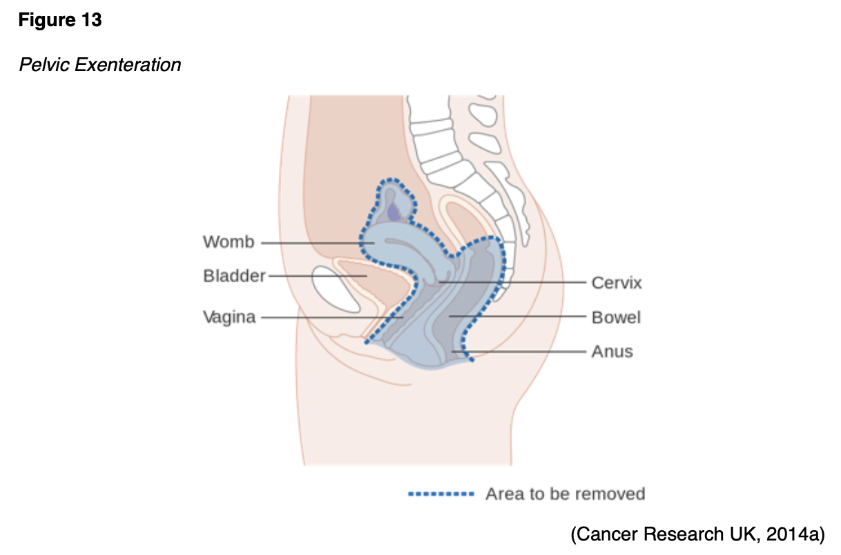
A complete parametrectomy/upper vaginectomy is a surgical procedure reserved for select patients with residual disease after hysterectomy. It involves surgical resection of the parametrium and removal of the upper portion of the vagina. It carries significant morbidity, including risks for bowel and bladder injury, incontinence, and sexual dysfunction. Surgery is not a therapeutic option for cervical cancers diagnosed at more advanced stages, such as stage IIB and higher (NCCN, 2021). However, surgery has a limited role in patients with persistent disease (continued evidence of cancer following definitive therapy) or localized cancer recurrence (cancer that returns in the pelvis only following a disease-free interval). Patients in either of these categories may be cured with a surgical procedure called pelvic exenteration. A pelvic exenteration is a drastic surgical procedure (see Figure 13) in which many, if not all, organs are removed from the pelvis. The extent of surgery varies but typically involves removing the uterus, fallopian tubes, ovaries, vagina, bladder, urethra, rectum, and anal sphincter. Following the procedure, the patient will have a permanent colostomy and a urinary diversion. A colostomy is formed by bringing the remaining intestine to the abdominal wall’s surface to provide an outlet (stoma) for stool to evacuate the bowel. With a urinary diversion, the kidneys and ureters are reconnected to a surgically created opening (urostomy) to drain the urine (NCCN, 2021; Yarbro et al., 2018).
Surgical Risks and Side Effects
The risks and side effects of surgery depend on the size and degree of cancer invasion, the extent of surgery, and the structures removed. All surgeries and invasive procedures for cervical cancer are accompanied by risks, such as adverse reactions to anesthesia, bleeding, blood clots, fistula formation (an abnormal connection between two hollow spaces within the body), bowel and bladder injury, infection, sexual dysfunction, and life-threatening sepsis. A loss of fertility can negatively impact interpersonal relationships, quality of life, psychological health, and emotional wellbeing. Patients who undergo pelvic exenteration have difficulty adapting to the sweeping life alterations from surgery and struggle to care for colostomy and urostomy devices. Similarly, patients may be affected by the physical and psychological aspects of body image distortion. The APRN serves a vital role in helping patients acclimate to these life-altering changes by facilitating healthy coping, addressing concerns, and referring patients to appropriate support groups or therapists (ACS, 2020a; NCCN, 2021; Yarbro et al., 2018).
Radiation Therapy
Radiation therapy is a type of localized treatment that delivers a precisely measured amount of high-energy, focused ionizing radiation to the tumor while causing as little injury as possible to the surrounding tissue. Radiation causes cellular damage to cancer cells, leading to biological changes in the DNA and rendering cells incapable of reproducing or spreading. All healthy cells and cancer cells are vulnerable to the effects of radiation and may be injured or destroyed; however, healthy cells can repair themselves and remain functional. The total dose of radiation is hyper-fractionated, which means it is delivered to the tumor in small, divided doses (i.e., fractions) rather than all at once. Hyper-fractionation gives healthy cells a chance to recover between treatments. The total number of fractions (doses) administered depends on the tumor size, location, reason for treatment, patient’s overall health, performance status, and goals of therapy, as well as any other therapies the patient is receiving. Radiation therapy plays a central role in treating cervical cancer and can be delivered externally or internally; some patients may receive both. The most common types of radiation used for cervical cancer include external beam radiation therapy (EBRT) and brachytherapy (ACS, 2020a; Nettina, 2019).
EBRT
EBRT delivers radiation from a source outside the body and is the most common type of radiation therapy used for cervical cancer. Traditionally, radiation beams could only match the tumor’s height and width, exposing more healthy tissue to the consequences of radiation. Further advancements in imaging technology have led to more precise treatment mechanisms that allow even more of the radiation beam to reach the tumor. Intensity-modulated radiation therapy (IMRT) is a newer, highly conformal form of radiation that offers modulation of the radiation beam’s intensity, delivering a higher radiation dose to a precise location, reducing unintended exposure to healthy tissues, enhancing clinical outcomes, and limiting side effects. In cervical cancer, IMRT helps minimize radiation exposure to the bowel and other critical structures, especially among patients who have undergone a hysterectomy. For cervical cancer, EBRT is most commonly administered concurrently with platinum-based chemotherapy to enhance the therapeutic response. The next section will discuss the role of chemoradiation (NCCN, 2021; Nettina, 2019).
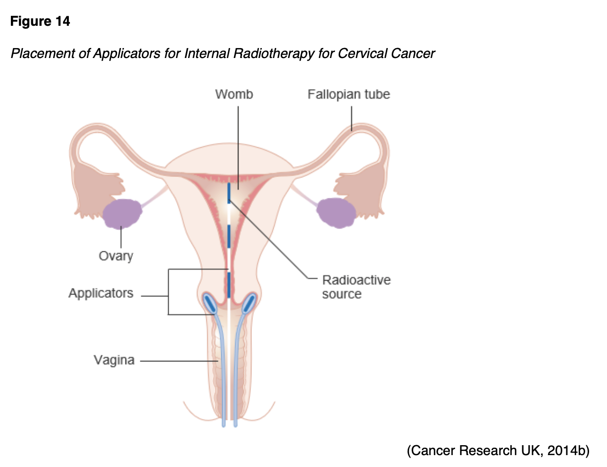
Brachytherapy
Brachytherapy is a critical component of cervical cancer treatment, particularly for patients who are not surgical candidates. It is also frequently used following EBRT to deliver an added boost of radiation to a specified area or to palliate symptoms. Brachytherapy’s intracavitary approach involves the internal implantation of an intrauterine device. The radioactive source is inserted directly into or near the tumor, as displayed in Figure 14.
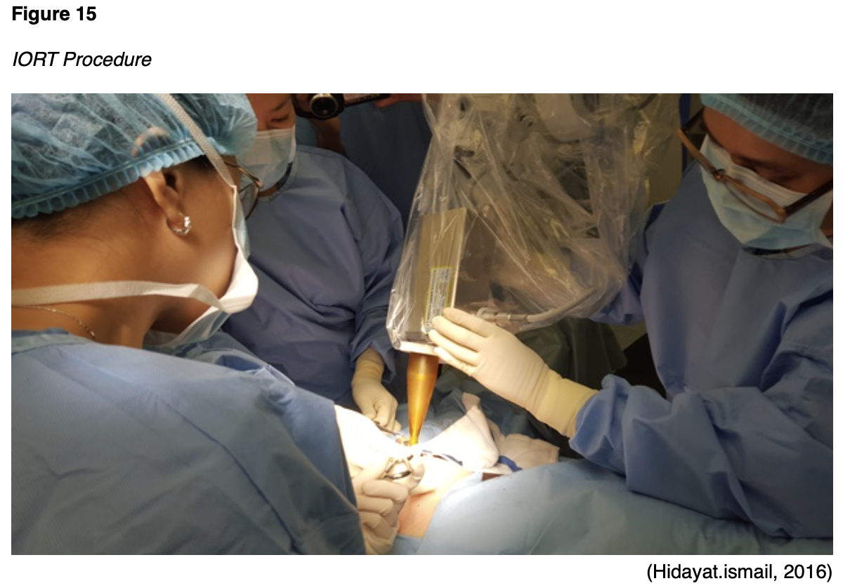
Intraoperative Radiation Therapy (IORT)
IORT is a specialized technique that delivers a single dose of highly focused radiation to the tumor bed during surgery (see Figure 15). IORT is particularly useful for patients with recurrent cancer within a previously irradiated field. During IORT, the overlying normal tissues and structures (e.g., bowel and surrounding organs) are manually displaced outside the radiation field to minimize exposure. IORT is delivered with electrons using preformed applicators matched to the surgically defined area of risk (NCCN, 2021).
Radiation Side Effects
Radiation side effects depend on the specific area(s) of the body exposed and the dose received. Superficial skin irritation at the site of the EBRT beams is likely and can include redness, blistering, and sunburn. The urinary system, bowel, and genitalia are most commonly affected by radiation for cervical cancer. Bladder dysfunction may manifest as dysuria, hematuria, acute kidney injury, hydronephrosis, or incontinence. Gastrointestinal (GI) issues are common due to the tumor’s anatomical location and the impact of radiation on surrounding tissues and structures. Patients may endure nausea, vomiting, diarrhea, constipation, blood in stools, pain with defecation, or a loss of anal sphincter control. Sexual dysfunction is likely, particularly dyspareunia, atrophic vaginitis (inflammation and dryness of the vaginal tissue), vaginal agglutination (fusion and fibrosis of the vaginal walls), and recurrent yeast infections. If the ovaries are within the radiation field, patients may experience a permanent loss of ovarian function. Systemic effects can include fatigue, weakness, and dehydration (Nettina, 2019; Yarbro et al., 2018).
Chemotherapy
Chemotherapy, also called cytotoxic or antineoplastic therapy, refers to a group of high-risk, hazardous medications that destroy cancer cells throughout the body. Chemotherapy works by interfering with the normal cell cycle, impairing DNA synthesis and cell replication to prevent cancer cells from dividing and multiplying (Yarbro et al., 2018). Chemotherapy serves a prominent role in the management of many types of cervical cancers. Platinum-based chemotherapy (e.g., cisplatin [Platinol]) with concurrent radiation (chemoradiation) is the standard of care for potentially curable cervical cancer that is not amenable to surgery. Cisplatin (Platinol) acts as a radiosensitizer, thereby rendering cancer cells more vulnerable to radiation’s toxic effects. According to the NCCN (2021), the best outcomes occur when chemoradiation is completed within an 8-week timeframe. Adjuvant chemotherapy (administered after surgery) aims to prevent cancer recurrence, reduce micro-metastases, and eradicate any remaining cancer cells. Chemotherapy also serves a proficient role in the treatment of recurrent cervical cancer and metastatic disease. In these instances, chemotherapy is considered palliative and can alleviate or delay cancer symptoms, enhance comfort, reduce symptom burden, improve quality of life, and extend survival. As noted in Table 6, systemic therapy regimens for recurrent or metastatic disease typically include intravenous formulations with at least two or three medications. There is a prominent role for bevacizumab (Avastin), a non-chemotherapy agent that is discussed in the next section (Itano, 2016; Olsen et al., 2019; NCCN, 2021).

Chemotherapy Side Effects
The side effects of chemotherapy vary based on the drug type, dosage, duration of treatment, and specific patient factors. Since cancer cells divide rapidly, chemotherapy is primed to target cells that divide rapidly, thereby impacting normal cells that divide quickly like those in the GI tract, skin/hair cells, and bone marrow. As a group, their most common side effects include lowered blood counts (anemia, thrombocytopenia, neutropenia), fatigue, nausea, anorexia, alopecia (hair loss), diarrhea, skin changes, and peripheral neuropathy (damage to the sensory nerves). Alopecia (hair loss) deserves special attention because it can cause significant emotional distress to women. Chemotherapy-induced hair loss generally begins with hair thinning, which occurs about 7-15 days after the first dose. This effect results from damage to the dividing hair matrix cells, which causes the hair shaft to break at the follicular orifice or hair bulb. While the degree of hair loss depends on the chemotherapy agent, dose, and administration schedule, paclitaxel (Taxol) and docetaxel (Taxotere) are well-known for inducing alopecia. APRNs should reassure women that their hair will begin to regrow within a few weeks following the cessation of chemotherapy, as permanent alopecia following chemotherapy is rare (Olsen et al., 2019).
Cisplatin (Platinol) is a moderate-to-highly emetogenic agent that induces both acute and delayed nausea. Poorly controlled chemotherapy-induced nausea and vomiting (CINV) are associated with unfavorable treatment compliance, impairing survival. Aprepitant (Emend) is approved to reduce CINV associated with cisplatin (Platinol) therapy. Available in oral and intravenous preparations, aprepitant (Emend) is a neurokinin-1 (NK-1) receptor antagonist that blocks substance P/neurokinin 1 in the brain. It is used in combination with a 5-hydroxytryptamine type 3 (5HT3) receptor antagonist (e.g., ondansetron [Zofran] or palonosetron [Aloxi]) and a corticosteroid (e.g., dexamethasone [Decadron]) for therapy (Aapro et al., 2015). Patients require aggressive hydration before and after the administration of cisplatin (Platinol) to manage associated nephrotoxicity and protect the renal system from injury (Brown et al., 2019; Olsen et al., 2019).
Chemotherapy-induced peripheral neuropathy (CIPN) is another common side effect of cisplatin (Platinol), carboplatin (Paraplatin), paclitaxel (Taxol), and docetaxel (Taxotere). It is often the dose-limiting toxicity (DLT) of these agents. DLTs are severe toxicities and side effects that are serious enough to warrant a dose reduction or discontinuation of the treatment. CIPN results from demyelination of the sensory and motor axons. Patients experience reduced nerve conduction velocity, leading to the loss of deep tendon reflexes, paresthesia (numbness and tingling), weakness, and burning pain. CIPN initially affects the body’s most distal points, such as the fingertips and toes, and moves proximally toward the midline as the damage progresses. In severe cases, patients may lose all sensation in the fingers, hands, toes, and feet; this can cause significant disability, such as the inability to grasp or hold items and gait disturbance, leading to imbalance and falls. CIPN is a complex topic since no single pathophysiologic process has been identified to explain the various neuropathies that occur following exposure to these chemotherapy agents. CIPN is dose-dependent and progressive during treatment but also can have a cascading effect after treatment ends. During this cascading phenomenon, symptoms become more prominent after discontinuation of the offending agent. Pain, sensory changes, and weakness that manifest during treatment generally lead to chemotherapy dose reductions, changes in treatment protocols, or termination of the therapeutic agent entirely. The morbidity associated with CIPN can lead to pronounced reductions in quality of life and independence with activities of daily living (Brown et al., 2019).
Currently, no medications or supplements are effective in preventing CIPN. Exercising regularly, reducing alcohol use, and treating preexisting medical conditions (vitamin B12 deficiency) may reduce the risk of CIPN. Management for CIPN is complex, and effective treatment options are limited. Pharmacologic treatment focuses on symptom relief, although many agents are not highly effective. Over-the-counter pain medications, menthol creams, capsaicin cream, or lidocaine patches may be used for comfort. Some patients may be prescribed medications such as gabapentin (Neurontin), an anticonvulsant/anti-epileptic agent that also can treat neuropathic pain. Some patients may find relief from selective serotonin-norepinephrine reuptake inhibitors (SNRIs) like duloxetine (Cymbalta). The American Society of Clinical Oncology (ASCO, 2020) recently released an updated statement on the management of CIPN, and new recommendations support the use of duloxetine (Cymbalta) as the only agent with appropriate evidence to support its use in patients with established painful CIPN, although the degree of benefit is limited (Loprinzi et al., 2020). Patients with CIPN must be counseled on ways to avoid injury through wearing supportive shoes and paying attention to home safety, such as using handrails on stairs and removing throw rugs. Patients must also be mindful of water temperatures, as they may become less sensitive to hot water, increasing their risk of burns when bathing or washing dishes. Improvement in function and resolution of symptoms often occur over time, but nerve damage may be permanent (Brown et al., 2019).
Hypersensitivity Reactions to Chemotherapy
A hypersensitivity reaction (HSR) occurs when the immune system becomes overstimulated by a foreign substance and creates antibodies, causing an immune response. HSRs are commonly associated with several chemotherapy agents that are used widely in cervical cancer treatment—most prominently paclitaxel (Taxol), docetaxel (Taxotere), and carboplatin (Paraplatin). HSR risk can be reduced by pre-medicating patients with corticosteroids, antihistamines, and/or acetaminophen (Tylenol). HSRs can occur during the initial chemotherapy infusion or after subsequent administrations of the same agent. Paclitaxel (Taxol) is well-known for its risk of nearly immediate acute HSR, whereas carboplatin (Paraplatin) more commonly induces an HSR after several cycles. The majority of HSRs occur during the first 15 minutes of the infusion. Initial signs and symptoms can include hives, urticaria, pruritis, swelling, back pain, facial flushing, rhinitis, abdominal cramping, chills, hypotension, and anxiety. Patients may require supplemental oxygen, fluid resuscitation, or other emergency medications as indicated. For life-threatening symptoms like bronchospasm, angioedema (swelling of the oral cavity, lips, or tongue), or anaphylaxis, epinephrine 0.1-0.5 mg (1:10,000 solution for adult patients) has been required (Nettina, 2019).
Targeted Therapy
Bevacizumab (Avastin) is a humanized monoclonal antibody that binds to and inhibits the activity of human vascular endothelial growth factor (VEGF) to its receptors, thereby blocking proliferation and formation of new blood vessels that supply tumor cells. VEGF is a signaling protein that stimulates angiogenesis (the formation of new blood vessels) in healthy and cancerous cells. Blood vessels carry oxygen and nutrients to the tissue, supporting growth and survival. Thus, tumors need blood vessels to grow and spread. Anti-angiogenesis is the process of inhibiting the formation of new blood vessels by blocking the VEGF receptors. Angiogenesis inhibitors (i.e., VEGF inhibitors) target the blood vessels that supply oxygen to the tumor cells, ultimately causing them to starve by cutting off their nutrient supply. VEGF inhibitors such as bevacizumab (Avastin) sever the blood supply to cancer cells by interfering with the VEGF receptor, so tumors stay small and eventually starve. Bevacizumab (Avastin) is commonly used as part of combination therapy to treat recurrent and metastatic cervical cancers. While it is generally well-tolerated, potential side effects include bleeding events, headaches, hypertension, and proteinuria (protein spilling in the urine due to increased pressure in the kidneys). Many patients require concurrent treatment with antihypertensives due to the medication’s elevation of blood pressure. Bevacizumab (Avastin) is contraindicated within 6 weeks of surgery (preoperatively or postoperatively) due to an increased risk for major bleeding events, delayed wound healing, and fistula formation. It also carries a black box warning for bowel perforation (a hole in the intestines), and patients should report any sudden onset of severe and diffuse abdominal pain, bloating, nausea, vomiting, or rectal bleeding (NCCN, 2021; Olsen et al., 2019).
Immunotherapy
Immunotherapy comprises a novel group of cancer treatments that stimulate the immune system to recognize and destroy cancer cells. Immunotherapy aims to produce antitumor effects by modifying the actions of the body’s natural host defense mechanisms, making them more sensitive to cancer cells. Immune-based treatments work differently than chemotherapy because they are highly specialized in their activity. Immune checkpoint inhibitors work by blocking the receptors that cancer cells use to inactivate immune cells (i.e., T-cells). When this signal is blocked, T-cells can better differentiate between healthy cells and cancer cells, thereby augmenting the cancer cells’ immune response. Checkpoint inhibitors occur in two categories: (1) programmed cell death-1 (PD-1)/PD-ligand 1 (PD-L1) inhibitors, and (2) cytotoxic T-lymphocyte–associated antigen 4 (CTLA-4) inhibitors. PD-1 is a transmembrane checkpoint protein expressed on the surface of circulating immune cells. PD-1 usually acts as an “off switch” to keep the immune cells from attacking other cells in the body. When PD-1 binds to PD-L1, it signals the T-cell to leave neighboring cells alone. Some cancer cells have large amounts of PD-L1, which helps them evade immune attack. PD-1 and PD-L1 inhibitors prevent this complex formation and enable immune cells to continue attacking tumor cells. Monoclonal antibodies that target PD-1 or PD-L1 can block this binding and boost the immune response against cancer cells (Sasikumar & Ramachandra, 2018).
The role of immunotherapy in the treatment of cervical cancers is less advanced than other diseases, and clinical research is ongoing. Currently, pembrolizumab (Keytruda) is the only agent used in this setting for metastatic cervical tumors that are MSI-H or dMMR. Pembrolizumab (Keytruda) is a humanized monoclonal antibody that binds with high affinity to PD-1, thereby preventing its interaction with PD-L1 and PD-L2. In the phase II KEYNOTE-158 clinical trial, Pembrolizumab (Keytruda) demonstrated promising and durable antitumor activity in patients with PD-L1-positive cervical cancer, offering a clinically meaningful and viable treatment strategy. Based on these results, the FDA granted accelerated approval of pembrolizumab (Keytryuda) for patients with advanced PD-L1–positive cervical cancer and disease progression or recurrent disease after chemotherapy. Pembrolizumab (Keytruda) is generally tolerated well; the most common side effects include fatigue, nausea, anorexia, coughing, diarrhea, skin rash, and itching. However, patients may experience severe and possibly fatal autoimmune-related adverse effects. Although any organ system can be affected, the most commonly observed reactions include colitis, hepatitis, endocrinopathies (thyroid and adrenals), pneumonitis, and rash progression to Stevens-Johnson syndrome (Chung et al., 2019; Feng et al., 2018; Sasikumar & Ramachandra, 2018).
HPV Vaccination
According to the World Health Organization (WHO, 2020), effective primary prevention (HPV vaccination) combined with secondary prevention strategies (screening and treating precancerous lesions) will thwart most cervical cancers. Cervical cancer is highly preventable, and one of the only two cancers that can be mitigated through screening; colorectal cancer is the other. Among women of all races, cervical cancer screening is lowest among women with no health insurance and women with lower education levels (CDC, 2019; 2020b). Three HPV vaccines have been approved by the FDA to protect against high-risk HPV subtypes that are linked to cancer: nine-valent (9vHPV, Gardasil 9), quadrivalent (4vHPV, Gardasil), and bivalent (2vHPV, Cervarix). Each offers protection against the high-risk oncogenic subtypes HPV-16 and HPV-18. Gardasil also protects against types 6 and 11, while Gardasil 9 further protects against types 31, 33, 45, 52, and 58. Gardasil was initially a quadrivalent vaccine offering protection against four different types of HPV; it was approved for use in 2006 for females aged 9 through 26 years. Initially, it was recommended as a series of three injections for females aged 11 through 26 years. In 2018, the FDA approved the expanded use of Gardasil 9—a 9-valent vaccine with increased coverage of additional high-risk HPV types—in those ages 9-45 years. Since the introduction of the HPV vaccine, HPV infections and cervical precancers have dropped significantly. Over the past 10 years, females aged 12-19 years have experienced a 71% decrease in high-risk HPV infections associated with cancer and anogenital warts. During the same timeframe, there has been a 61% decrease in high-risk HPV infections in females aged 30-45 years. Precancerous cervical lesions caused by HPV types 16 and 18 have dropped by 40% among vaccinated women. Clinical data demonstrate that vaccination protection remains high after 10 years, with no decrease in protection when measuring antibodies in the blood. Still, nearly one-half (50%) of all adolescent girls have not been vaccinated against HPV in the United States (CDC, 2019; 2020b; Meites et al., 2019; NCI, 2019a; Siegel et al., 2020).
Side Effects
According to the CDC (2019), more than 100,000,000 doses of HPV vaccinations have been administered in the United States over the past 10 years, and vaccination is considered highly safe. Some of the most commonly reported side effects include the following:
- pain, redness, or edema near the injection site;
- fever;
- dizziness or fainting immediately after the injection;
- nausea;
- headaches or tiredness; and
- joint or muscle pain (CDC, 2019; NCI, 2019a).
Schedule and Dosing
The CDC’s Advisory Committee on Immunization Practices (ACIP) develops recommendations regarding all vaccination schedules and regimens in the United States. The current ACIP recommendations for HPV vaccination are as follows:
Routine or catch-up vaccination
- HPV vaccination is routinely recommended at age 11-12 years, but vaccination may begin as early as 9 years.
- HPV vaccination is recommended for all individuals up to 26 years who were not adequately vaccinated.
- The 2- or 3-dose series depends on the patient’s age at initial vaccination:
- Age 9 through 14 years at initial vaccination: follow the 2-dose series and administer injections at 0 and 6-12 months
- the minimum interval between vaccinations is 5 months
- repeat dose if administered too soon
- Age 15 years or older at initial vaccination: following the 3-dose series and administer injections at 0, 1-2 months, and 6 months
- the minimum interval between vaccinations is as follows:
- dose 1 to dose 2: 4 weeks
- dose 2 to dose 3: 12 weeks
- dose 1 to dose 3: 5 months
- repeat dose if administered too soon
- the minimum interval between vaccinations is as follows:
- Age 27 through 45 years: The ACIP recommends shared clinical decision-making for patients aged 27 through 45 years who have not previously been vaccinated to discuss the risk for new HPV infections and the possible benefits of vaccination with their health care provider. HPV vaccination in this age range provides less benefit because more people have already been exposed to the virus but has still demonstrated efficacy. Adults who were not vaccinated against HPV should discuss the risk for new HPV infections and the possible benefits of vaccination with their health care provider to determine if vaccination is still advised.
- Age 9 through 14 years at initial vaccination: follow the 2-dose series and administer injections at 0 and 6-12 months
Special Circumstances
- Immunocompromised patients, including those with HIV infection: administer the 3-dose series outlined above
- Children with a history of sexual abuse or assault: initiate at age 9 years
- Pregnancy: delay HPV vaccination until after pregnancy, but pregnancy testing is not required before vaccination. There is no evidence that vaccination will affect pregnancy or harm a fetus (CDC, 2020b; Meites et al., 2019).
Learn more about HPV vaccination with our HPV NursingCE course and earn 2.0 ANCC contact hours.
References
Aapro, M., Carides, A., Rapoport, B. L., Schmoll, H., Zhang, L., & Warr, D. (2015). Aprepitant and fosaprepitant: A 10-year review of efficacy and safety. The Oncologist, 20, 450-458. https://www.ncbi.nlm.nih.gov/pmc/articles/PMC4391760/pdf/theoncologist_14229.pdf
American Cancer Society. (2019). Cancer facts & figures for African Americans 2019-2021. https://www.cancer.org/content/dam/cancer-org/research/cancer-facts-and-statistics/cancer-facts-and-figures-for-african-americans/cancer-facts-and-figures-for-african-americans-2019-2021.pdf
American Cancer Society. (2020a). Cervical cancer. https://www.cancer.org/cancer/cervical-cancer.html
American Cancer Society. (2020b). Survival rates for cervical cancer. https://www.cancer.org/cancer/cervical-cancer/detection-diagnosis-staging/survival.html
American Cancer Society. (2020c). Tests for cervical cancer. https://www.cancer.org/cancer/cervical-cancer/detection-diagnosis-staging/how-diagnosed.html
American Cancer Society. (2020d). The American cancer society guidelines for the prevention and early detection of cervical cancer. https://www.cancer.org/cancer/cervical-cancer/detection-diagnosis-staging/cervical-cancer-screening-guidelines.html
American Cancer Society. (2020e). The HPV test. https://www.cancer.org/cancer/cervical-cancer/detection-diagnosis-staging/screening-tests/hpv-test.html
American Cancer Society. (2020f). What is cervical cancer? https://www.cancer.org/cancer/cervical-cancer/about/what-is-cervical-cancer.html
The American College of Obstetricians and Gynecologists. (2020). Loop electrosurgical excision procedure [LEEP]. https://www.acog.org/en/Womens%20Health/FAQs/Loop%20Electrosurgical%20Excision%20Procedure
Arbyn, M., Snijders, P. J. F., Meijer, C. J. L. M., Berkoff, J., Cuschieri, K., Kocjan, B. J., & Poljak, M. (2015). Which high-risk HPV assays fulfill criteria for use in primary cervical cancer screening? Clinical Microbiology and Infection, 21(9), 817-826. https://doi.org/10.1016/j.cmi.2015.04.015
Balasubramaniam, S. D., Balakrishnan, V., Oon, C. E., & Kaur, G. (2019). Progression of CIN to cancer [image]. https://commons.wikimedia.org/wiki/File:HpvInfectedSquamousEpithelialCell.png
Bhatla, N., Aoki, D., Sharma, D. N., & Sankaranarayanan, R. (2018). Cancer of cervix uteri. International Journal of Gynecology & Obstetrics, 143(Suppl. 2), 22-36. https://doi.org/10.1002/ijgo.12611
Brossel, R. (2017). The development of cervical cancer [image]. https://commons.wikimedia.org/wiki/File:The_development_of_cervical_cancer.jpg
Brown, T. J., Sedhom, R., & Gupta, A. (2019). Chemotherapy-induced peripheral neuropathy. JAMA Oncology, 5(5),750. https://doi.org/10.1001/jamaoncol.2018.6771
BruceBlaus. (2015). Radical hysterectomy [image]. https://commons.wikimedia.org/wiki/File:Hysterectomy.png
BruceBlaus. (2016). Cervical cryotherapy [image]. https://commons.wikimedia.org/wiki/File:Cervical_Cryotherapy.png
BruceBlaus. (2017). LEEP [image]. https://commons.wikimedia.org/wiki/File:LEEP.png
Bruni, L., Albero, G., Serrano, B., Mena, M., Gomez, D., Munoz, J. Bosch, F., de Sanjose, S. (2019). Human papillomavirus and related diseases report: United States of America. HPV Information Centre, 1-125. https://hpvcentre.net/statistics/reports/USA.pdf
Burness, J. V., Schroeder, J. M., & Warren, J. B. (2020). Cervical colposcopy: Indications and risk assessment. American Family Physician, 102(1), 39-48. https://www.aafp.org/afp/2020/0701/p39.html
Cancer Research UK. (2014a). Pelvic exenteration [image]. https://commons.wikimedia.org/wiki/File:Diagram_showing_the_area_removed_with_a_posterior_exenteration_for_cancer_of_the_cervix_CRUK_288.svg
Cancer Research UK. (2014b). Placement of applicators for internal radiotherapy for cervical cancer [image]. https://commons.wikimedia.org/wiki/File:Diagram_showing_the_position_of_the_applicators_for_internal_radiotherapy_for_cervical_cancer_CRUK_344.svg
Cancer Research UK. (2014c). Radical trachelectomy [image]. https://commons.wikimedia.org/wiki/File:Diagram_showing_the_parts_removed_with_trachelectomy_surgery_CRUK_338.svg
CancerQuest. (2020). Cancer genes. https://www.cancerquest.org/about-
us/contact-cancerquest
Cancer Statistics Center. (2020). Cervix. https://cancerstatisticscenter.cancer.org/#!/cancer-site/Cervix
Centers for Disease Control and Prevention. (2019). Human papillomavirus (HPV). https://www.cdc.gov/hpv/index.html
Centers for Disease Control and Prevention. (2020a). Basic information about HPV and cancer. https://www.cdc.gov/cancer/hpv/basic_info/index.htm
Centers for Disease Control and Prevention. (2020b). Immunization schedules for health care providers. https://www.cdc.gov/vaccines/schedules/index.html
Chung, H. C., Ros, W., Delord, J., Perets, R., Italiano, A., Shapiro-Frommer, R.,
Manzuk, L., Piha-Paul, S. A., Xu, L., Zeigenfuss, S., Pruitt, S. K., & Leary, A. (2019). Efficacy and safety of pembrolizumab in previously treated advanced cervical cancer: Results from the phase II KEYNOTE-158 study. Journal of Clinical Oncology, 37(17), 1470-1478. https://doi.org/10.1200/JCO.18.01265
Fang, J., Yu, X., Zhang, S., & Yang, Y. (2018). Effect of smoking on high-grade cervical cancer in women on the basis of human papillomavirus infection studies. Journal of Cancer Research and Therapeutics, 14(8), S184-S189. https://doi.org/10.4103/0973-1482.179190
Feng, Y., Ji, W., Yue, N., Huang, Y., & Ma, X. (2018). The relationship between PD-1/PD-L1 pathway and DNA mismatch repair in cervical cancer and its clinical significance. Cancer Management and Research, 10, 105-113. https://doi.org/ 10.2147/CMAR.S152232
Häggström, M. (2020). Positioning of SCJ and TZ [image].
https://commons.wikimedia.org/wiki/File:Transformation_zone_types
.svg
Hahn, A. W., & Spach, D. H. (2018). Human papillomavirus infection. https://www.std.uw.edu/go/pathogen-based/hpv/core-
concept/all
Hidayat.ismail. (2016). IORT procedure [image].
https://commons.wikimedia.org/wiki/File:IORT_procedure.jpg
Hodgson, A., & Park, K. J. (2019). Cervical adenocarcinomas: A heterogeneous group of tumors with variable etiologies and clinical outcomes. Archives of Pathology & Laboratory Medicine, 143(1), 34-46. https://doi.org/10.5858/arpa.2018-0259-RA
Khan, M. J., Werner, C. L., Darragh, T. M., Guido, R. S., Mathews, C., Moscicki, A.,
Mitchell, M. M., Schiffman, M., Wentzensen, N., Massad, L., Stewart, M., Mayeaux, E. J., Waxman, A. G., Conageski, C., Einstein, M. H., & Huh, W. K. (2017). ASCCP colposcopy standards: Role of colposcopy, benefits, potential harms, and terminology for colposcopic practice. Journal of Lower Genital Tract Disease, 21(4), 223-229. https://doi.org/10.1097/lgt.0000000000000338
Loprinzi, C. L., Lacchetti, C., Bleeker, J., Cavaletti, G., Chauhan, C., Hertz, D. L., Kelley, M. R., Lavino, A., Lustberg, M. B., Paice, J. A., Schneider, B. P., Smith, E. M. L., Smith, M. L., Smith, T. J., Wagner-Johnston, N., & Hershman, D. L. (2020). Prevention and management of chemotherapy-induced peripheral neuropathy in survivors of adult cancers: ASCO guideline update. Journal of Clinical Oncology, 38(28), 3325-3348. https://doi.org/10.1200/JCO.20.01399
Mehta, S., & Sachdeva, P. (2017). Colposcopy of female genital tract. Springer.
Meites, E., Szilagyi, P. G., Chesson, H. W., Unger, E. R., Romero, J. R., & Markowitz, L. E. (2019). Human papillomavirus vaccination for adults: Updated recommendations of the advisory committee on immunization practices. Morbidity and Mortality Weekly Report (MMWR), 68(32), 698-702. http://dx.doi.org/10.15585/mmwr.mm6832a3
Mello, V., & Sundstrom, R. K. (2020). Cervical intraepithelial neoplasia. StatPearls [Internet]. https://www.ncbi.nlm.nih.gov/books/NBK544371/
Mysid. (2006). Anatomical location of the internal and external os [image]. https://commons.wikimedia.org/wiki/File:Gray1167.svg
Nakamura, M., Obata, T., Daikoku, T., & Fujiwara, H. (2019). The association and significance of p53 in gynecologic cancers: The potential of targeted therapy. International Journal of Molecular Sciences, 20(5482), 1-16. https://doi.org/10.3390/ijms20215482
National Cancer Institute. (2019a). Human papillomavirus (HPV) vaccines. Retrieved from https://www.cancer.gov/about-cancer/causes-prevention/risk/infectious-agents/hpv-vaccine-fact-sheet
National Cancer Institute. (2019b). Understanding cervical changes: Next steps after an abnormal screening test. https://www.cancer.gov/types/cervical/understanding-cervical-changes#2
National Comprehensive Cancer Network. (2021). NCCN clinical practice guidelines in oncology (NCCN guidelines®): Cervical cancer version 1.2021 - October 2, 2020. https://www.nccn.org/professionals/physician_gls/pdf/cervical.pdf
Nettina, S. M. (2019). Lippincott manual of nursing practice (11th ed.). Wolters Kluwer.
Nicolas, I., Saco, A., Barnadas, E., Marimon, L., Rakislova, N., Fuste, P., Rovirosa, A., Gaba, L., Bunesch, L., Gil-Ibanez, B., Pahisa, J., Dias-Feijoo, B., Torne, A., Ordi, J., & del Pino, M. (2020). Prognostic implications of genotyping and p16 immunostaining in HPV-positive tumors of the uterine cervix. Modern Pathology, 33, 128-137. https://doi.org/10.1038/s41379-019-0360-3
Olsen, M., LeFebvre, K., & Brassil, K. (2019). Chemotherapy and immunotherapy guidelines and recommendations for practice (1st ed.). Oncology Nursing Society.
Pal, A., & Kundu, R. (2020). Human papillomavirus E6 and E7: The cervical cancer hallmarks and targets for therapy. Frontiers in Microbiology, 10(3116). https://doi.org/10.3389/fmicb.2019.03116
Perkins, R. B., Guido, R. S., Castle, P. E., Chelmow, D., Einstein, M. H., Garcia, F., Huh, W. K., Kim, J. J., Moscicki, A., Nayar, R., Saraiya, M., Sawaya, G. F., Wentzensen, N., & Schiffman, M. (2020). 2019 ASCCP risk-based management consensus guidelines for abnormal cervical cancer screening tests and cancer precursors. Journal of Lower Genitourinary Tract Disease, 24(2), 102-131. https://doi.org/10.1097/LGT.0000000000000525
Sasikumar, P. G., & Ramachandra, M. (2018). Small-molecule immune checkpoint inhibitors targeting PD-1/PDL1 and other emerging checkpoint pathways. BioDrugs, 35(5), 481-497. https://doi.org/10.1007/s40259-018-0303-4.
Siegel, R. L., Miller, K. D., & Jemal, A. (2020). Cancer statistics, 2020. CA Cancer Journal for Clinicians, 70, 7-30. https://doi.org/10.3322/caac.21590
Tempfer, C. B., Tischoff, I., Dogan, A., Hilal, Z., Schultheis, B., Kern, P., & Reziczek, G. A. (2018). Neuroendocrine carcinoma of the cervix: A systematic review of the literature. BMC Cancer, 18(530), 1-16. https://doi.org/10.1186/s12885-018-4447-x
Teoh, D., Musa, F., Salani, R., Huh, W., & Jimenez, E. (2020). Diagnosis and management of adenocarcinoma in situ: A society of gynecologic oncology evidence-based review and recommendations. Obstetrics & Gynecology, 135(4), 869-878. https://doi.org/10.1097/AOG.0000000000003761
Truth Initiative. (2019). Women & tobacco. https://truthinitiative.org/sites/default/files/media/files/2019/03/Truth_Women%20and%20Tobacco_FactSheet_final.pdf
U.S. National Library of Medicine. (2020). TP53 gene: Tumor protein p53. https://medlineplus.gov/genetics/gene/tp53/#conditions
Wang, X., Huang, X., & Zhang, Y. (2018). Involvement of human papillomaviruses in cervical cancer. Frontiers in Microbiology, 9 (2896), 1-14. https://doi.org/10.3389/fmicb.2018.02896
World Health Organization. (2019). Human papillomavirus (HPV) and cervical cancer. https://www.who.int/news-room/fact-sheets/detail/human-papillomavirus-(hpv)-and-cervical-cancer
World Health Organization. (2020). Cervical cancer. https://www.who.int/health-topics/cervical-cancer#tab=tab_1
Wright, J. D., Goff, B., & Chakrabarti, A. (2020). Cervical intraepithelial neoplasia: Management. UpToDate. https://www.uptodate.com/contents/cervical-intraepithelial-neoplasia-management
Yarbro, C. H., Wujcik, D., & Gobel, B. H. (Eds.). (2018). Cancer nursing: Principles and practice (8th ed.). Jones & Bartlett Learning.
Zebitay, A. G., Gungor, E. S., IIhan, G., Cetin, O., Dane, C., Furtuna, C., Atmaca, F. F., & Tuna, M. (2017). Cervical conization and the risk of premature birth: A population-based multicentric trial of Turkish cohort. Journal of Clinical & Diagnostic Research, 11(3), QC21-QC24. https://doi.org/10.7860/JCDR/2017/22996.9495