About this course:
The purpose of this module is to provide an overview of cervical precancer and cervical cancer, outlining the most common risk factors, signs and symptoms, and treatment modalities to inform nursing practice and improve patient outcomes.
Course preview
The purpose of this module is to provide an overview of cervical precancer and cervical cancer, outlining the most common risk factors, signs and symptoms, and treatment modalities to inform nursing practice and improve patient outcomes.
By the completion of this learning activity, the nurse should be able to:
- Discuss the epidemiology of cervical cancer in the US and risk factors for the development of the disease
- Discuss the anatomy of the cervix and the pathophysiology leading to the development of cervical precancer and cancer
- Summarize the types of cervical cancer screening tests and the impact of human papillomavirus (HPV) on the development of the condition, and the role of the HPV vaccine in cervical cancer prevention
- Identify the signs and symptoms of cervical cancer, and clinical features as critical components of cervical cancer staging
- Describe the management of cervical precancer and invasive cervical cancer, including an overview of treatment risks, side effects, and the key aspects of patient education
According to the American Cancer Society (ACS, 2020a), approximately 13,800 women in the United States will be diagnosed with invasive cervical cancer in 2020, and about 4,290 will die from the disease. Previously cited as one of the most common causes of cancer-related deaths, cervical cancer mortality rates have declined dramatically over the past 30 years with the advent and widespread utilization of the Papanicolaou (Pap) smear, followed by the addition of the HPV co-test. The recent arrival of the HPV vaccination has ignited a new era in cervical cancer prevention, demonstrating a substantial reduction in invasive cervical cancer since its inception. When diagnosed early and managed effectively, cervical cancer is highly treatable (ACS, 2020a; Bruni et al., 2019).
Epidemiology
Cervical cancer is the fourth most common cancer among women, with 0.6% of women (1 in 159) diagnosed during their lifetime (Siegel et al., 2020). According to the World Health Organization (WHO, 2020), the annual age-adjusted incidence rate of cervical cancer is 7.6 per 100,000 people. Cervical cancer incidence is 30% higher in non-Hispanic Black (NHB) women than non-Hispanic White women (NHW; ACS, 2019). The states with the highest annual incidence and mortality rates include California (1,630 diagnoses/510 deaths), Texas (1,410/430), Florida (1,130/360), and New York (930/260), respectively (Cancer Statistics Center, 2020). The average age of diagnosis is 50, and the disease occurs most frequently in women aged 35 to 44 years. While 20% of cases occur in women over the age of 65, cervical cancer rarely develops in those who have maintained compliance with routine cervical cancer screenings (ACS, 2020a). Overall, cervical cancer ranks as the 12th leading cause of female cancer deaths in the United States; it is the second leading cause of cancer deaths in women aged 20 to 39 years (Siegel et al., 2020) and the third leading cause of cancer deaths in women aged 15 to 44 years (Bruni et al., 2019). The 5-year survival rate of localized cervical cancer (i.e., cancer that has not spread outside of the cervix or uterus) is 92%, declining to 17% for those with distant metastases (i.e., cancer spreading to organs or body parts outside of the cervix or uterus). NHB women are more likely to be diagnosed at advanced stages of the disease, reducing their 5-year survival rate to 56% compared to 68% in NHW women (ACS, 2019; 2020b). While survival has improved since the 1970s for most cancer types, it has remained relatively stagnant for cervical cancers; this is attributed to the lack of significant treatment advances for patients with recurrent and metastatic disease. Furthermore, nearly 90% of women who die from cervical cancer have inadequate access to prevention, screening, and treatment (WHO, 2020).
Risk Factors
Risk factors for cervical cancer include HPV infection, tobacco use, early onset of sexual activity, multiple sexual partners, and HIV infection. Predominantly transmitted through sexual contact, HPV is the chief risk factor for cervical cancer, leading to nearly 99% of diagnoses. Tobacco use is the only nonsexual behavior associated with cervical dysplasia (abnormal growth of cells on the cervix’s surface) and cancer, serving as a significant cofactor for the development of HPV. Women who smoke are at least twice as likely as non-smokers to be diagnosed with cervical cancer. Research has demonstrated that among women with HPV infections, those who smoke have a significantly higher viral load on the cervix, heightening the risk of cervical cancer development (Fang et al., 2018; Truth Initiative, 2019). Early sexual activity and multiple sexual partners (especially those who have had numerous partners themselves) increase the risk of HPV exposure. People who are immunocompromised—such as those with HIV, transplant recipients, cancer patients, and those receiving immunosuppressive medications—are at higher risk for acquiring an HPV infection (ACS, 2020a).
Pathophysiology
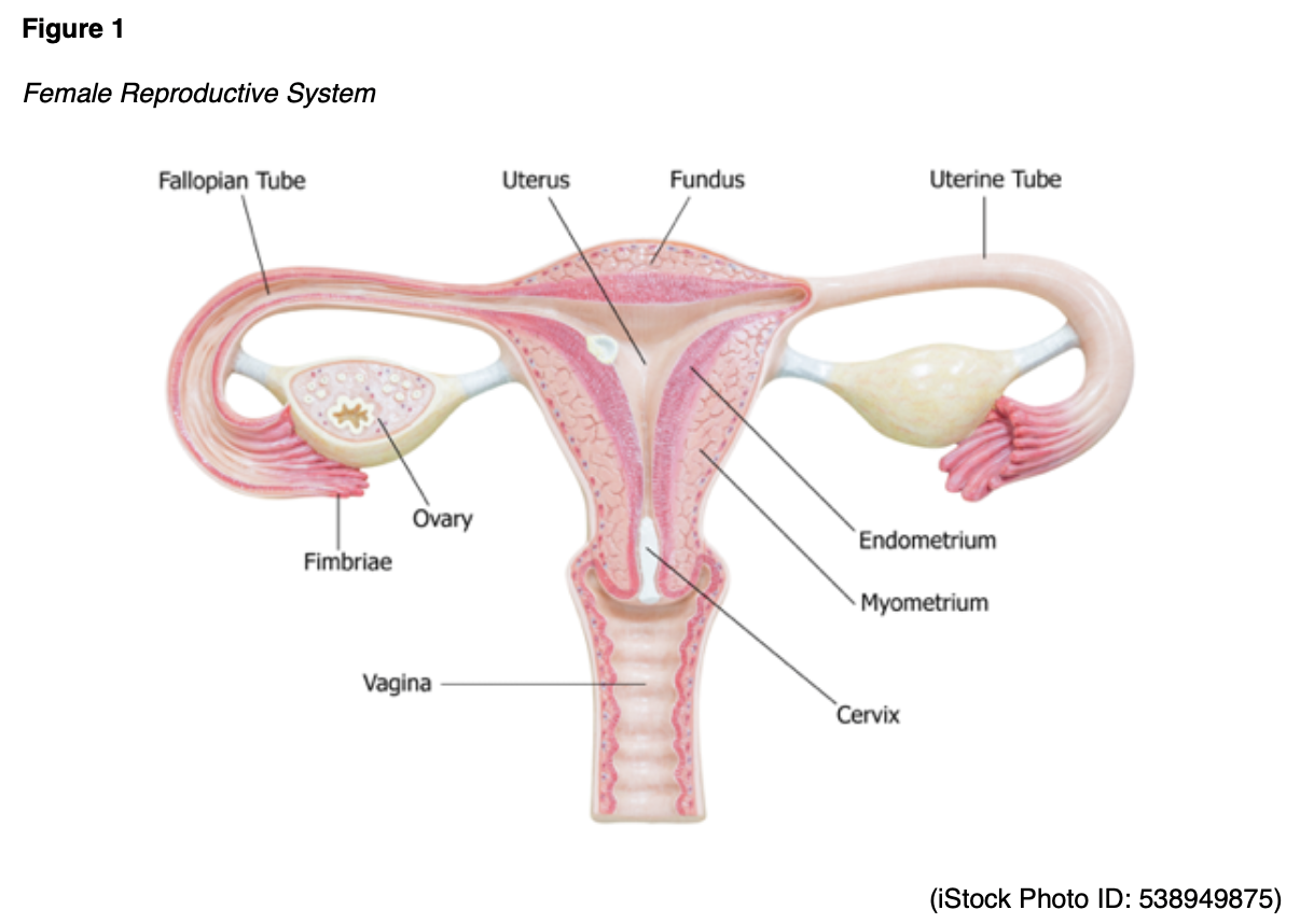
The cervix is the fibromuscular lower portion of the uterus (the hollow, pear-shaped organ that carries a fetus) that connects the uterine cavity to the vagina, as demonstrated in Figure 1. It measures 4 cm in length and 3 cm in diameter in an adult woman of childbearing age and undergoes progressive involution following menopause. The cervix is substantially larger in parous women (i.e., those who have given birth) than nulliparous women (i.e., those who have never carried a pregnancy to term; McCance & Heuther, 2019; Mehta & Sachdeva, 2017).
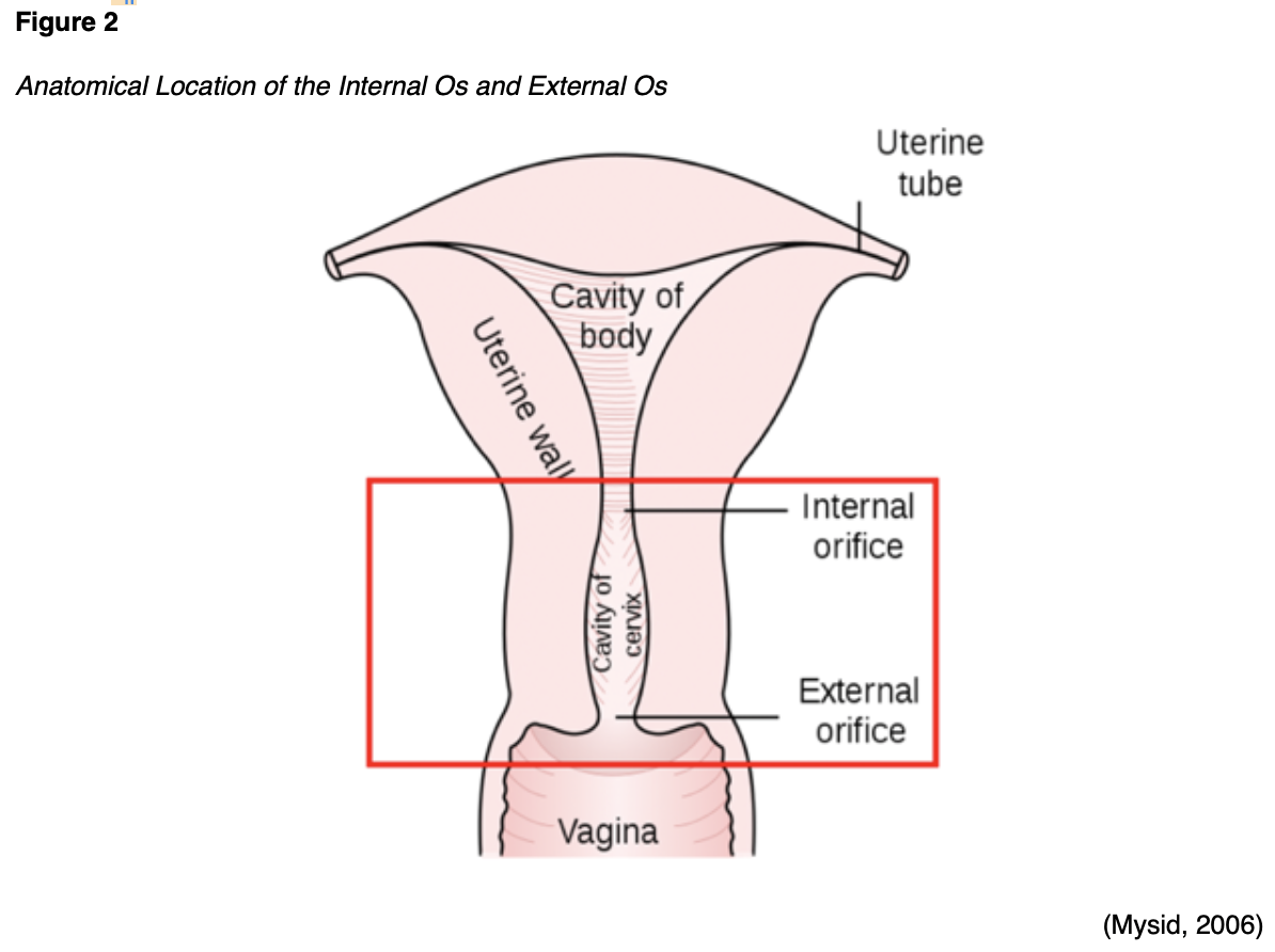
Two sections comprise the cervix: the ectocervix and the endocervix. The ectocervix extends into the vagina and is lined with stratified squamous non-keratinized epithelium. The squamous epithelium involves four layers: basal, parabasal, intermediate, and superficial. As demonstrated in Figure 2, the cervix opens into the vagina through the external os (external orifice), which is the readily visible portion of the cervix. The external os marks the transition point to the endocervix (endocervical canal), which is the inner part of the cervix lined by mucus-secreting columnar epithelium or glandular cells. The endocervix is rarely visible on an internal pelvic examination. It forms a passageway between the external and internal os (internal orifice or isthmus). The internal os is a narrow opening that marks the transition from the endocervix to the endometrium, which is the lining of the uterine cavity. The cervix enables the passage of sperm into the uterine cavity through dilation of the external and internal os. The cervix maintains a sterile environment by frequent shedding of the endometrium, thick cervical mucus production, and a narrow external os to protect against bacterial invasion (McCance & Heuther, 2019; Mehta & Sachdeva, 2017).
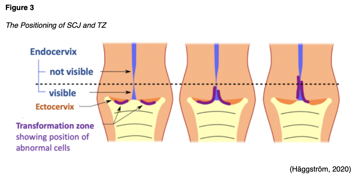
The area between the endocervix and the ectocervix is called the squamocolumnar junction (SCJ) and is where cervical dysplasia occurs most often. At birth, the SCJ lies close to the external os, but its location shifts with age, parity, and hormonal status, moving internally. Figure 3 displays the variable positions of the SCJ. The columnar epithelium inside the cervix expands outward onto the ectocervix, exposing the tissue to estrogen and irritants from the vagina’s acidic environment. Over time, the columnar epithelium is replaced with metaplastic squamous epithelium, forming the new SCJ. Metaplasia is a process by which one mature cell type transforms into another mature cell type that is not naturally present in that l
...purchase below to continue the course
HPV
HPV is a ubiquitous group of tiny viruses that carry deoxyribonucleic acid (DNA) that can adapt to their host and evade the immune system. An estimated 12,000 new cases of HPV-associated cervical cancers are diagnosed in the United States each year (Centers for Disease Control and Prevention [CDC], 2020b). A growing body of evidence links HPV to other anogenital cancers (anal, vulvar, vaginal, penile), as well as head and neck cancers (Bruni et al., 2019). Of the 200 identified strains of HPV, approximately 40 have a particular affinity for the genital region. While most of these are low-risk and non-oncogenic (wart-causing), at least 13 are high-risk and oncogenic (cancer-causing), leading to cervical cancer (CDC, 2020a; Wang et al., 2018). The most common drivers of cervical carcinogenesis are the high-risk subtypes HPV-16 and HPV-18, which are responsible for at least 70% of cervical cancers (Bruni et al., 2019; WHO, 2019). The majority of sexually active males and females will be infected with HPV at some point during their lives, but most of these infections will remain clinically silent. The immune system often mediates the spontaneous clearance of HPV from the cervix within 2 years of acquiring the infection. However, about 10% of women with HPV will develop a persistent infection (Mello & Sundstrom, 2020). Chronic infections with high-risk HPV subtypes can lead to cervical precancers or cervical intraepithelial neoplasia (CIN; CDC, 2020a; Hahn & Spach, 2018).
The Role of HPV in Cervical Carcinogenesis
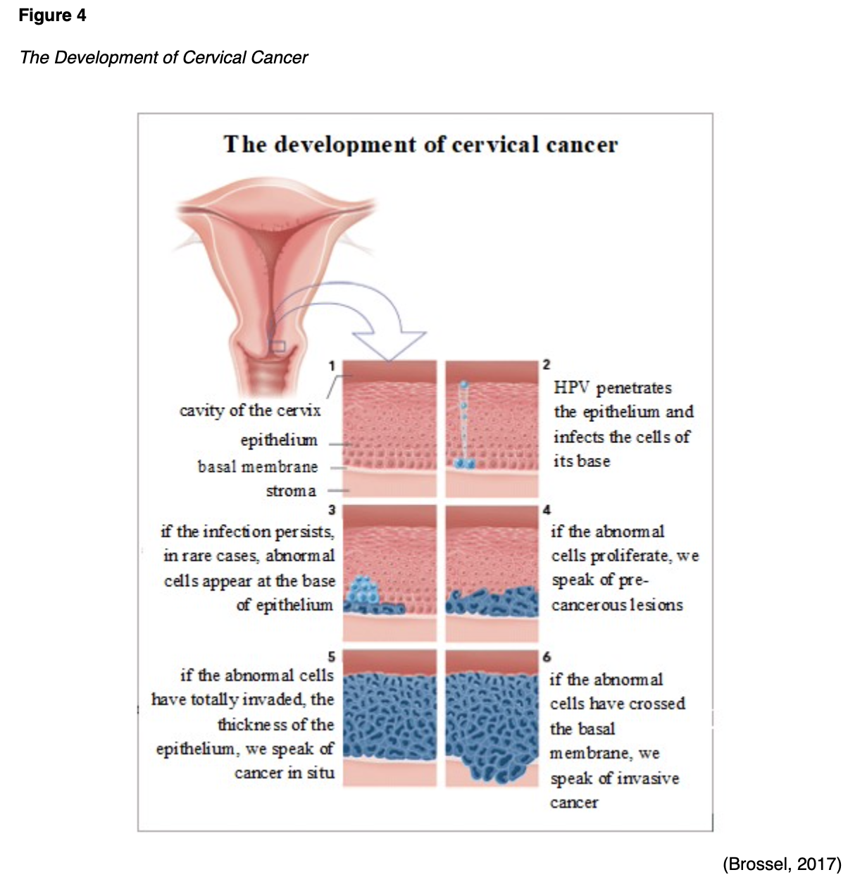
HPV leads to CIN and invasive cancer through a series of pathologic processes in which oncogenes and tumor suppressor genes serve fundamental roles. An oncogene is a mutation associated with cancer development; it can be inherited or caused by exposure to carcinogenic substances in the environment, such as ultraviolet (UV) radiation and free radicals. In their normal and non-mutated state, oncogenes are called proto-oncogenes, regulating healthy cell growth and division. When a proto-oncogene mutates into an oncogene, it becomes permanently activated (turned on), fueling unregulated cell growth and resultant cancer cells. Tumor suppressor genes are healthy genes that slow cell division, repair DNA errors, and induce apoptosis (programmed cell death). Under physiologic conditions, tumor suppressor genes regulate healthy cellular growth and division and prevent cells with mutated or damaged DNA from replicating. When these tumor suppressor genes are inactivated (turned off), these processes become unregulated, thereby fueling cancer growth. The malignant transformation process and the role of HPV are demonstrated in Figure 4 (CancerQuest, 2020; Hahn & Spach, 2018; Pal & Kundu, 2020).
Pap Smear and HPV Testing
Since HPV-induced CIN does not cause symptoms, cellular changes are identified on screening Pap smear and HPV testing. A Pap smear is a noninvasive test to evaluate for cervical abnormalities, precancerous lesions, and invasive cancer. The Pap smear does not diagnose HPV itself and requires specific HPV testing. In the US, HPV testing is most commonly performed as a co-test alongside the Pap smear. HPV can be detected about a year after infection in the cervix. The US Food & Drug Administration (FDA) has approved several HPV tests, all of which evaluate the cells’ internal environment explicitly for signs of infection with high-risk viral subtypes. Viral nucleic acid (DNA or mRNA) must be detected within the cervical cells to form a definitive diagnosis. Nurses provide patient-centered care across the continuum of healthcare and serve crucial roles in disease prevention and health promotion. Nurses can have a tremendous impact on cervical cancer prevention by ensuring patients receive accurate information to make informed decisions about screening. While there are variations in cervical cancer screening guidelines, the ACS (2020e) recommends that women aged 25 to 65 years undergo HPV testing with a Pap smear every 5 years. The interval for follow-up cervical cancer screening depends on the results and individual patient factors as dictated by the treating clinician (ACS, 2020d; 2020e; Perkins et al., 2020).
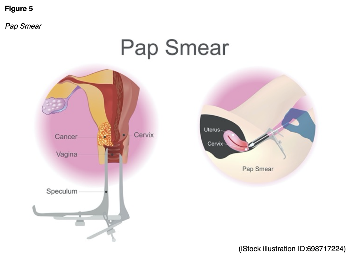
As shown in Figure 5, a Pap smear is a simple, outpatient test in which a vaginal speculum is placed inside the vagina to visualize the cervix. The cervix is scraped to collect cervical cells using a small, cone-shaped brush and plastic spatula. Next, the brush and spatula are rinsed in a liquid-filled vial and sent to the laboratory for processing. Nurses can provide reassurance to patients that the test is well-tolerated and rarely causes complications or pain. Mild vaginal spotting may occur immediately following the Pap smear, which resolves in less than 24 hours (National Cancer Institute [NCI], 2019b).
Pap Smear Results
The results of the Pap smear denote whether the cervical cells are normal or abnormal, but some samples may return as unsatisfactory (see below). All Pap smears must include cells within the SCJ to be satisfactory. Since nurses are routinely tasked with communicating results to patients, it is essential to ascertain a foundational understanding of the significance of Pap smear results to accurately educate patients and respond to questions (Yarbro et al., 2018).
Normal Results
Normal (negative) results indicate no abnormal cells were identified, and patients do not require further workup or management.
Unsatisfactory Results
Unsatisfactory Pap smear results indicate that there were insufficient cells to evaluate the specimen definitively, or the cells may have been obscured by blood or mucus. Patients with unsatisfactory results typically require repeat testing.
Abnormal Results
Abnormal (positive) results indicate abnormal cellular changes in the sample and any grade of dysplasia warrants further workup. There are several types of abnormal Pap results; the most common include the following:
- Atypical squamous cells of undetermined significance (ASCUS)
- ASCUS is the most common abnormal Pap result and is an inconclusive finding that does not determine the origin of the abnormal cells or whether the changes are secondary to an HPV infection. ASCUS can be caused by benign conditions, and it typically requires a repeat Pap smear in 1 year.
- Atypical glandular cells (AGC)
- AGC signifies that some glandular cells do not appear normal.
- Squamous intraepithelial lesion (SIL)
- SIL refers to dysplastic changes in the cervix and is categorized into low-grade (LSIL) or high-grade (HSIL). LSIL denotes early cellular changes (mild dysplasia), meaning cells have only a few abnormal characteristics and still resemble healthy cells. HSIL denotes moderate or severe dysplasia, in which cells appear highly abnormal under the microscope but remain confined to the cervix’s surface. While HSIL does not indicate invasion into deeper parts of the cervix, it can develop into cervical cancer if it is not treated.
- Carcinoma in situ (CIS)/ Adenocarcinoma in situ (AIS)
- CIS denotes early-stage noninvasive cancer, in which the cancerous cells arise from and are limited to the squamous epithelium. CIS has not penetrated more deeply into the tissues.
- AIS indicates a noninvasive lesion of the glandular tissue and is much less common than CIS. AIS has not penetrated more deeply into the tissues.
- Invasive cervical cancer
- Less commonly, Pap smears can detect malignant cells; however, invasive cervical cancer is rarely identified on a Pap smear in women who have undergone screening at regular intervals, as cervical abnormalities typically manifest as precancerous lesions (ACS, 2019; NCI, 2019b; Mello & Sundstrom, 2020; Perkins et al., 2020).
HPV Test Results
An HPV test result is either positive (high-risk HPV was found) or negative (high-risk HPV was not found; Arbyn et al., 2015; Perkins et al., 2020).
Follow-Up of Abnormal Results
While the treating clinician will develop the follow-up plan for abnormal results, nurses are often involved in educating patients on the procedures, managing expectations, and answering questions. This section provides an overview of the most common procedures performed in follow-up to abnormal Pap smear or HPV results.
Colposcopy
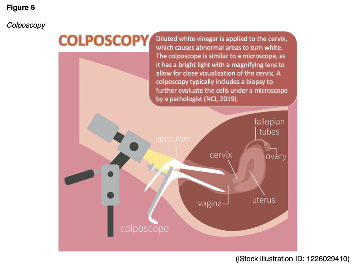
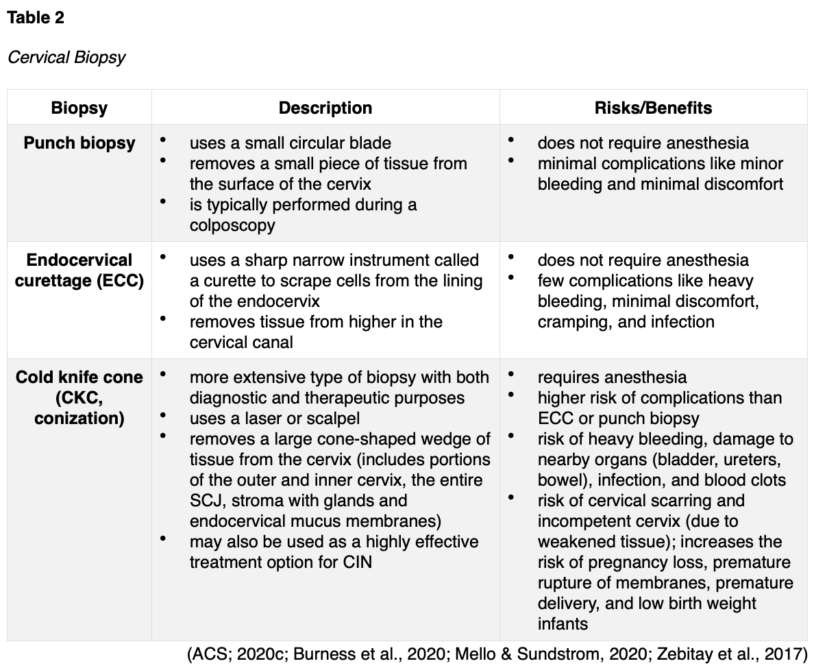
A colposcopy (see Figure 6) is a noninvasive procedure in which a weak acetic acid solution (diluted vinegar) is applied to the cervix. The vinegar turns abnormal areas white, allowing for direct visualization of cervical changes under a stereomicroscope. A biopsy of the abnormal area is performed, as tissue sampling and histological analysis are the only way to diagnose precancer or cancer definitively. There are three major types of cervical biopsies, as outlined in Table 2. Nurses can help reassure patients that colposcopy is a safe procedure that carries minimal risks. The potential harms may include discomfort, psychological distress, infection, and bleeding (ACS, 2020c; Burness et al., 2020; Khan et al., 2017).
Cervical Intraepithelial Neoplasia (CIN)
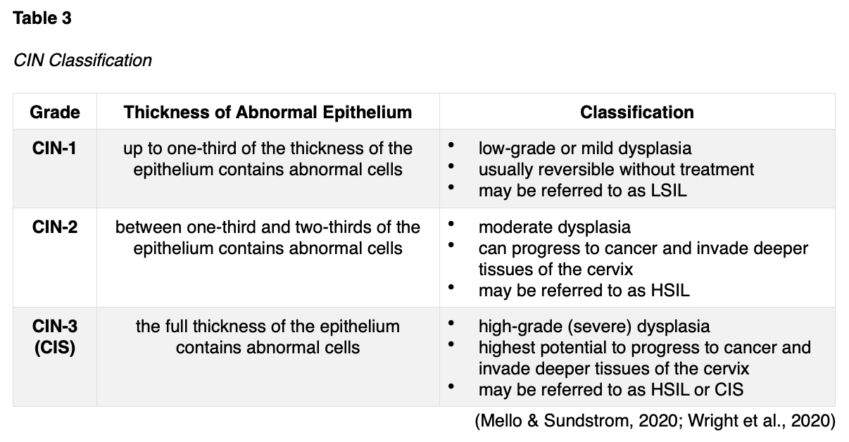
CIN is a histological diagnosis determined from a biopsy specimen. CIN is a noninvasive, precancerous condition in which the abnormal cells are confined to the epithelium and relates directly to the integration of an HPV infection. It denotes the extent of dysplasia within the layers of the tissue and is classified into three grades based on how deeply the abnormal cells invade into the skin. As the CIN grade advances, the risk for progression to invasive cancer increases. The classification of CIN is further delineated in Table 3. Figure 7 depicts the process by which early changes (CIN-1) progressively affect the deeper layers of the epithelium, leading to full-thickness involvement in its most severe form (CIN-3/CIS; Mello & Sundstrom, 2020).

CIN Management and Treatment
CIN treatment aims to eradicate the precancerous lesions to prevent them from progressing to invasive cancer. Treatment selection depends on various factors such as the extent and severity of the CIN, the patient’s age, their medical history, cost-effectiveness, availability, and the potential risks and benefits of each method. Procedures to treat CIN can affect a woman’s future childbearing potential, so shared decision-making is crucial to ensure patients are fully informed of the potential risks before deciding on a therapy option. Most low-grade CINs resolve without intervention; only about 1% of cases will progress to cervical cancer. While most patients with CIN-1 can be adequately managed with close observation using Pap smears, HPV testing, and colposcopies as indicated, patients with high-grade CIN typically require intervention. Treatment options include ablative and excisional techniques. Ablative techniques (e.g., cryotherapy) destroy abnormal tissues by freezing them, whereas excisional methods (e.g., loop electrosurgical excision procedure [LEEP] and CKC, described in Table 1) remove the abnormal tissue surgically. Since LEEP and CKC are the two most common and effective excisional procedures for CIN-2, CIN-3, and CIS, it is important that nurses understand these procedures to provide effective patient education (Perkins et al., 2020; Wright et al., 2020).
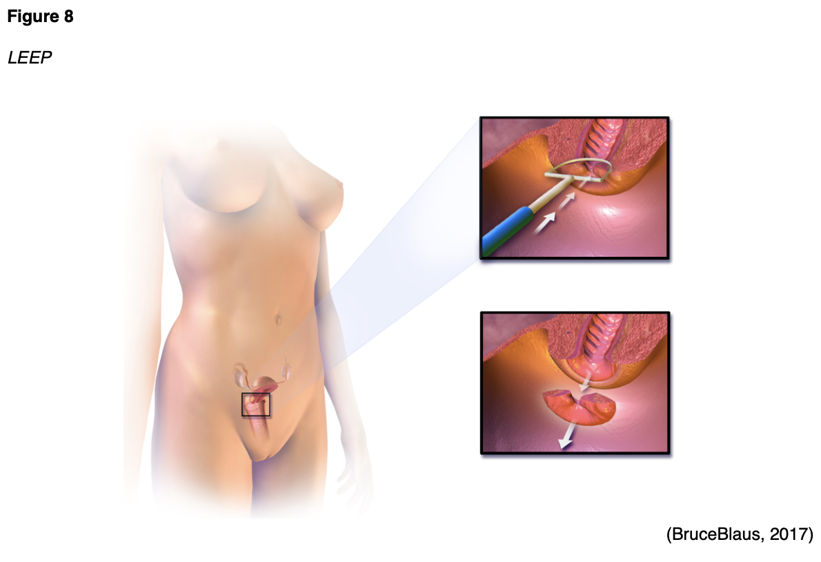
Loop Electrosurgical Excision Procedure (LEEP)
LEEP is a widely utilized treatment for high-grade cervical dysplasia following colposcopy. As shown in Figure 8, a thin wire loop that functions as a scalpel is used to excise abnormal cells from the cervix. An electric current is passed through the loop to remove a thin layer of the cervix. Nurses can prepare patients that local anesthesia is used for pain control so they may only feel minimal discomfort during the procedure, if at all. The procedure is usually performed in the outpatient setting and only takes about 15 minutes. At the end of the procedure, a reddish-brown paste called ferric subsulfate (Monsel’s solution) may be applied to the cervix to control any bleeding; alternatively, the blood vessels may be cauterized (heated with a device to control bleeding). If ferric subsulfate (Monsel’s solution) is used, a harmless brownish vaginal discharge may occur for up to 48 hours after the procedure. The most common side effects include mild discomfort and slight bleeding within the first 2 to 3 weeks after the procedure. Patients should be advised to avoid placing anything inside the vagina (including tampons and douching) to allow the cervix to heal fully. Similarly, they should abstain from sexual intercourse for 4 to 6 weeks. Patients may shower as usual but should avoid baths. A LEEP is associated with a small increased risk for future pregnancy complications, including premature birth and low birth weight. In rare cases, the cervix may narrow after the procedure, causing menstrual irregularities (American College of Obstetricians and Gynecologists, 2020; Perkins et al., 2020).
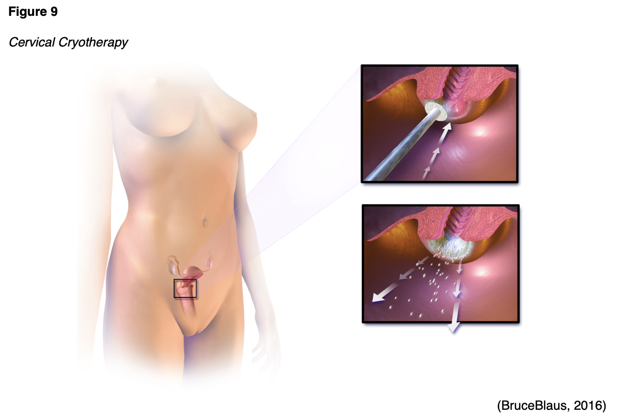
Cryotherapy
Cryotherapy, which is also referred to as cryosurgery or laser ablation, is demonstrated in Figure 9. It involves applying a cooled metal disc (cryoprobe) to the cervix to freeze the precancerous cells. The cryoprobe is cooled using compressed carbon dioxide (CO2) or nitrous oxide (NO) gas. Cryotherapy is a simple, fast outpatient procedure that is performed without anesthesia. It typically takes about 15 minutes and may cause mild discomfort. Following cryotherapy, the frozen area regenerates with normal epithelium in about a month, and some patients can experience watery vaginal discharge during this time. Patients should avoid sexual intercourse or placing anything in the vagina during the healing process for 4 to 6 weeks (Mello & Sundstrom, 2020; Perkins et al., 2020).
Cervical Cancer Subtypes
Cervical cancer ensues when precancerous cells invade the basement membrane. It occurs in two major subtypes: squamous cell carcinoma (SCC) and adenocarcinoma. The majority of cervical cancers (>80%) are SCC, originate in the squamous epithelium in the TZ, and closely correlate with chronic HPV infection as described earlier. Cervical adenocarcinoma begins in the glandular cells that line the upper portion of the cervix and cause up to 20% of cervical cancers. Adenocarcinomas originate in the TZ or the glandular epithelium above the TZ and are distinct from SCC, as they are not uniformly caused by infection with HPV (Bruni et al., 2019; Hodgson & Park, 2019). Neuroendocrine carcinoma of the cervix (NECC) is a third, rare, and more aggressive type that accounts for only 1.5% of all cervical cancers. There are four subtypes of NECC tumors: small cell neuroendocrine carcinoma (SCNEC), large cell neuroendocrine carcinoma (LCNEC), typical carcinoid tumor, and atypical carcinoid tumor. Of these, SCNEC is the most common and aggressive type and carries the poorest prognosis. In contrast, carcinoid tumors typically have a more indolent disease trajectory and a more favorable prognosis. The role of HPV in mediating the development of NECC tumors is not clearly understood. Unlike SCC and adenocarcinoma, NECC is not universally preceded by preinvasive disease. Therefore, early detection on Pap smear screening is less common. The management of NECC is more complex and less precise due to the rarity of the condition but typically includes multiple chemotherapy agents (Tempfer et al., 2018).
Signs and Symptoms
Women with early-stage cervical cancers typically do not have symptoms. With cervical cancer, symptoms do not develop until the cancer advances, grows larger, and invades nearby tissue. The most common symptoms include the following:
- abnormal vaginal bleeding, such as spotting between periods, bleeding after intercourse, menstrual periods that are longer or heavier than usual, and postmenopausal bleeding;
- dyspareunia (pain during or immediately following intercourse) or pain in the pelvis not related to intercourse;
- abnormal vaginal discharge;
- swelling of the legs due to impaired lymphatics caused by tumor compression;
- urinary symptoms such as frequency, dysuria, inability to void, or hematuria; and
- constipation (ACS, 2020a).
Principles of Pathologic Review
The biopsy sample undergoes a series of tests to determine the cancer’s pathologic features, evaluate its behavior, and select the best treatment options. Tumor grade is a measurement of how different the cancer cells look from healthy cells under the microscope. The grading determines the likelihood of cancer growth and spread and is defined as follows:
- Grade 1 is well-differentiated (appears similar to healthy cells) and the least aggressive.
- Grade 2 is moderately differentiated, appears less like healthy cells, and is an intermediate grade (more aggressive than grade 1).
- Grade 3 is poorly differentiated or undifferentiated (does not resemble healthy cells at all), is high-grade and most aggressive, and tends to grow and spread more quickly (ACS, 2020a; Yarbro et al., 2018).
Cancer Staging
Worldwide, the staging system of the International Federation of Gynecology and Obstetrics (FIGO) guides cervical cancer staging. The guidelines were last updated in 2018 and are integrated within the National Comprehensive Cancer Network (NCCN) guidelines. The four stages of cervical cancer are displayed in Figure 10. Since many cases require specialized surgical care, all patients with suspected cervical cancers should be promptly referred to a gynecologic oncologist (ACS, 2020b; Bhatla et al., 2018; NCCN, 2021).

Cervical Cancer Treatment
The most optimal cervical cancer treatment depends on various factors, such as the pathologic features, cancer stage, plans for fertility, patient preference, age, and medical history. Treatment is often multimodal, with several therapies combined and administered simultaneously (concurrently) or sequentially. This section will provide a synopsis of the most common evidence-based treatment strategies (ACS, 2020a; NCCN, 2021).
Surgery
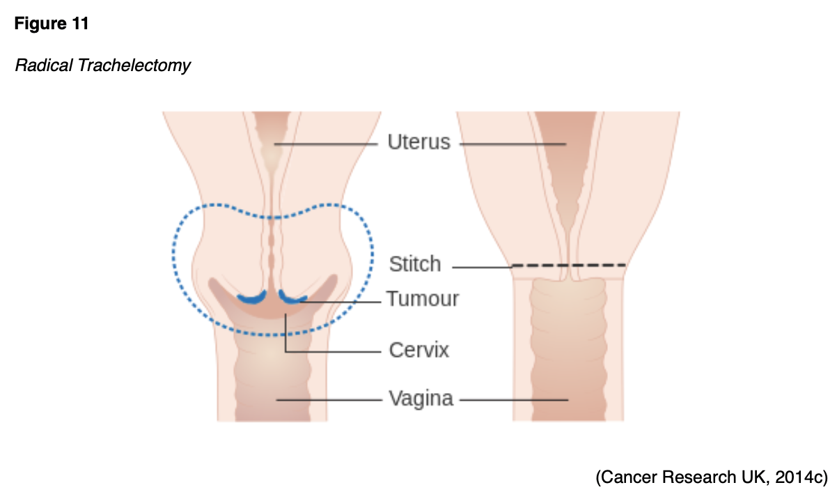
Surgical intervention for cervical cancer can be fertility-sparing or non-fertility-sparing. Most early-stage cervical cancers can be effectively managed with LEEP or CKC, both of which are fertility-sparing treatments. A radical trachelectomy, also called a cervicectomy, involves the surgical removal of the cervix, upper vagina, and supporting ligaments, but the uterine corpus is preserved (see Figure 11). The surrounding pelvic lymph nodes are also removed to evaluate for cancer spread. With regard to its impact on future pregnancies, a radical trachelectomy is associated with a 10% likelihood of second-trimester loss, but more than 70% of women carry gestation to 37 weeks or more (NCCN, 2021).
When fertility preservation is not desired or irrelevant, early-stage cervical cancers are managed with radical hysterectomy (see Figure 12). Patients with early-stage cervical cancer may undergo a radical hysterectomy with bilateral pelvic lymphadenectomy or pelvic lymph node dissection (PLND). A radical hysterectomy involves removing the uterus, cervix, a portion of the vagina, and the parametrium (the connective tissue that surrounds the cervix). PLND refers to the removal of the lymph nodes from the pelvis; the ovaries and fallopian tubes remain intact (NCCN, 2021).

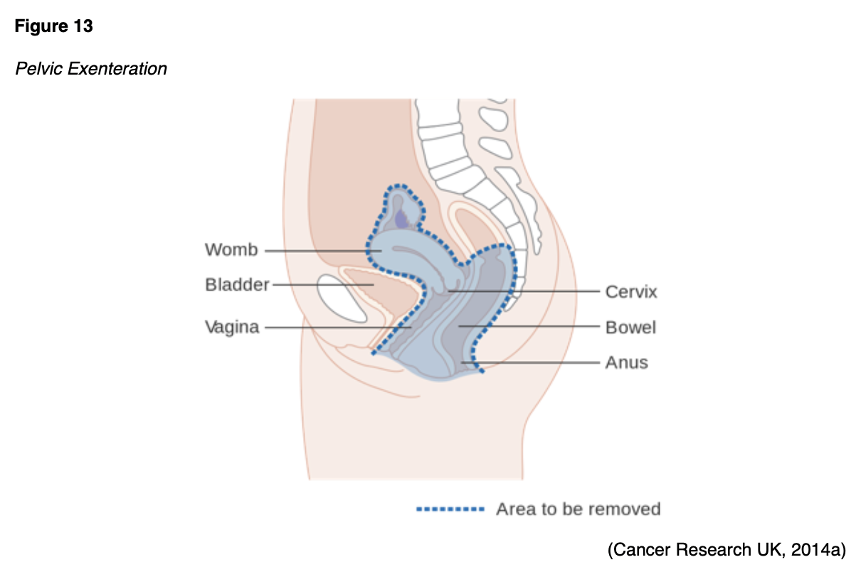
A complete parametrectomy/upper vaginectomy is a surgical procedure reserved for select patients with residual cancer following a hysterectomy. It involves surgical resection of the parametrium and removal of the upper portion of the vagina. It carries significant morbidity, including risks for bowel and bladder injury, incontinence, and sexual dysfunction. Less commonly, some patients may undergo a drastic surgical procedure called a pelvic exenteration (see Figure 13), in which many, if not all, the organs are removed from the pelvis. The extent of the surgery varies but typically involves removing the uterus, fallopian tubes, ovaries, vagina, bladder, urethra, rectum, and anal sphincter. Following the procedure, the patient will have a permanent colostomy and a urinary diversion. A colostomy is formed by bringing the remaining intestine to the abdominal wall’s surface to provide an outlet (stoma) for stool to evacuate the bowel. With a urinary diversion, the kidneys and ureters are reconnected to a surgically created opening (urostomy) to drain the urine (NCCN, 2021; Yarbro et al., 2018).
Surgical Risks and Side Effects
The risks and side effects of surgery depend on the size and degree of cancer invasion, the extent of surgery, and the structures removed. All surgeries and invasive procedures for cervical cancer are accompanied by risks, such as adverse reactions to anesthesia, bleeding, blood clots, fistula formation (an abnormal connection between two hollow spaces within the body), bowel and bladder injury, infection, sexual dysfunction, and life-threatening sepsis. Patients who undergo pelvic exenteration often have difficulty adapting to the sweeping life alterations from surgery and struggle to care for colostomy and urostomy devices. Patients may have complications with newly formed ostomies, such as malfunctioning of the stoma, skin breakdown, and leaking at the site (Yarbro et al., 2018). Nurses serve a vital role in helping patients acclimate to the physical care of their new stoma and ostomy device, which can pose extensive challenges. Patients are often affected by the physical and psychological aspects of body image distortion and lifestyle adjustments. Studies have demonstrated that nurses are key in identifying and meeting patients’ psychosocial needs and managing negative emotions toward ostomy care. Compassionate and knowledgeable nursing care focused on managing psychosocial needs, reducing anxiety, and addressing depression is helpful in alleviating patients’ stress responses and promotes healthy coping and adjustment (Jin et al., 2019). Further, a loss of fertility can negatively impact interpersonal relationships, quality of life, psychological health, and emotional wellbeing. Nurses are tasked with addressing these issues with sensitivity, empathy, and compassion, fostering a safe and non-judgmental environment for patients to openly express their concerns. Nurses can offer psychosocial support and connect patients with resources, support groups, and medical specialists (Yarbro et al., 2018).
Radiation Therapy
Radiation therapy is a type of localized treatment that delivers a precisely measured amount of high-energy, focused ionizing radiation to the tumor while causing as little injury as possible to the surrounding tissue. Radiation causes cellular damage to cancer cells, leading to biological changes in the DNA and rendering cells incapable of reproducing or spreading. All healthy cells and cancer cells are vulnerable to the effects of radiation and may be injured or destroyed; however, healthy cells can repair themselves and remain functional. The total dose of radiation is hyper-fractionated, which means it is delivered to the tumor in small, divided doses (i.e., fractions) rather than all at once. Hyper-fractionation gives healthy cells a chance to recover between treatments. Radiation therapy plays a central role in treating cervical cancer and can be delivered externally or internally; some patients may receive both. The most common types of radiation used for cervical cancer include external beam radiation therapy (EBRT) and brachytherapy (ACS, 2020a; Nettina, 2019).
EBRT
EBRT delivers radiation from a source outside the body and is the most common type of radiation therapy used for cervical cancer. Traditionally, radiation beams could only match the tumor’s height and width, exposing more healthy tissue to the consequences of radiation. Further advancements in imaging technology have led to more precise treatment mechanisms that allow even more of the radiation beam to reach the tumor. Intensity-modulated radiation therapy (IMRT) is a newer, highly conformal form of radiation that offers modulation of the radiation beam’s intensity, delivering a higher radiation dose to a precise location, reducing unintended exposure to healthy tissues, enhancing clinical outcomes, and limiting side effects. In cervical cancer, IMRT helps minimize radiation exposure to the bowel and other critical structures, especially among patients who have undergone a hysterectomy. For cervical cancer, EBRT is most commonly administered concurrently with platinum-based chemotherapy to enhance the therapeutic response. The next section will discuss the role of chemoradiation (NCCN, 2021; Nettina, 2019).
Brachytherapy
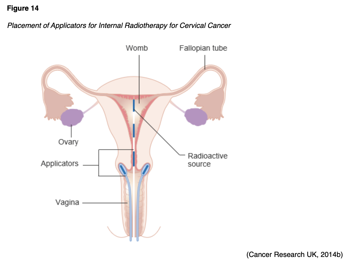
Brachytherapy is a critical component of cervical cancer treatment, particularly for patients who are not surgical candidates. It is also frequently used following EBRT to deliver an added boost of radiation to a specified area or to palliate symptoms. Brachytherapy’s intracavitary approach involves the internal implantation of an intrauterine device. The radioactive source is inserted directly into or near the tumor, as displayed in Figure 14.
Intraoperative Radiation Therapy (IORT)
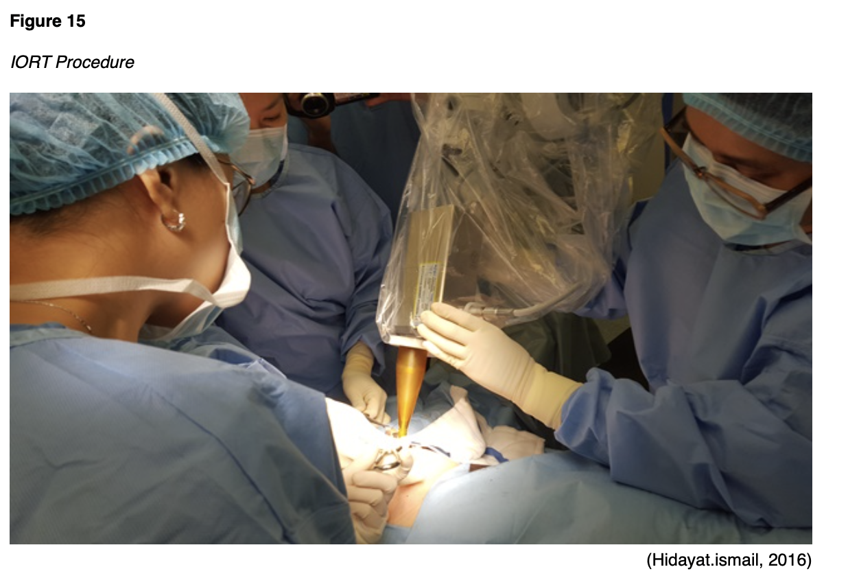
IORT is a specialized technique that delivers a single dose of highly focused radiation to the tumor bed during surgery (see Figure 15). IORT is particularly useful for patients with recurrent cancer within a previously irradiated field. During IORT, the overlying normal tissues and structures (e.g., bowel and surrounding organs) are manually displaced outside the radiation field to minimize exposure. IORT is delivered with electrons using preformed applicators matched to the surgically defined area of risk (NCCN, 2021).
Radiation Side Effects
Radiation side effects depend on the specific area(s) of the body exposed and the dose received. Superficial skin irritation at the site of the EBRT beams is likely and can include redness, blistering, and sunburn. The urinary system, bowel, and genitalia are most commonly affected by radiation for cervical cancer. Bladder dysfunction may manifest as dysuria, hematuria, acute kidney injury, hydronephrosis, or incontinence. Gastrointestinal (GI) issues are common due to the tumor’s anatomical location and the impact of radiation on surrounding tissues and structures. Patients may endure nausea, vomiting, diarrhea, constipation, blood in stools, pain with defecation, or a loss of anal sphincter control. Sexual dysfunction is likely, particularly dyspareunia, atrophic vaginitis (inflammation and dryness of the vaginal tissue), vaginal agglutination (fusion and fibrosis of the vaginal walls), and recurrent yeast infections. If the ovaries are within the radiation field, patients may experience a permanent loss of ovarian function. Systemic effects can include fatigue, weakness, and dehydration (Nettina, 2019; Yarbro et al., 2018).
Chemotherapy
Chemotherapy, also called cytotoxic or antineoplastic therapy, refers to a group of high-risk, hazardous medications that destroy cancer cells throughout the body. Chemotherapy works by interfering with the normal cell cycle, impairing DNA synthesis and cell replication to prevent cancer cells from dividing and multiplying (Yarbro et al., 2018). Chemotherapy serves a prominent role in the management of many types of cervical cancers. Platinum-based chemotherapy (e.g., cisplatin [Platinol]) with concurrent radiation (chemoradiation) is the standard of care for potentially curable cervical cancer that is not amenable to surgery. Cisplatin (Platinol) acts as a radiosensitizer, thereby rendering cancer cells more vulnerable to radiation’s toxic effects. Adjuvant chemotherapy (administered after surgery) aims to prevent cancer recurrence, reduce micro-metastases, and eradicate any remaining cancer cells. Chemotherapy also serves a proficient role in the treatment of recurrent cervical cancer and metastatic disease. In these instances, chemotherapy is considered palliative and can alleviate or delay cancer symptoms, enhance comfort, reduce symptom burden, improve quality of life, and extend survival (Olsen et al., 2019; NCCN, 2021).
Chemotherapy Side Effects
Side effects of chemotherapy are inevitable due to the nonspecific nature of cytotoxic therapy; it simultaneously impacts healthy cells along with cancerous cells. However, side effects vary based on the drug type, dosage, duration of treatment, and specific patient factors. Not all patients respond in the same way, and not all chemotherapy agents pose the same risks. Assessment and education are the most critical components to ensuring timely recognition, intervention, and management of side effects as experienced by each patient. Many side effects, such as nausea, can be thwarted by implementing appropriate prevention strategies and medications. As a group, the most common side effects include bone marrow suppression (anemia, thrombocytopenia, neutropenia), fatigue, anorexia, mucositis (mouth sores), nausea, vomiting, diarrhea, and skin changes, and alopecia (hair loss). Alopecia deserves special attention because it can cause significant emotional distress to women. Chemotherapy-induced hair loss generally begins with hair thinning, which occurs about 7-15 days after the first dose. This effect results from damage to the dividing hair matrix cells, which causes the hair shaft to break at the follicular orifice or hair bulb. While the degree of hair loss depends on the chemotherapy agent, dose, and administration schedule, paclitaxel (Taxol) and docetaxel (Taxotere) are well-known for inducing alopecia. Nurses should reassure patients that their hair typically begins to regrow within a few weeks following the cessation of chemotherapy, as permanent alopecia following chemotherapy is rare (Nettina, 2019; Olsen et al., 2019).
Cisplatin (Platinol) is a moderate-to-highly emetogenic agent that induces both acute and delayed nausea. Poorly controlled chemotherapy-induced nausea and vomiting (CINV) are associated with unfavorable treatment compliance, impairing survival. Nurses are essential to ensuring nausea is well-managed by appropriately pre-medicating with anti-emetics and educating patients regarding how to take anti-emetic medications at home. Further, patients also require aggressive hydration before and after the administration of cisplatin (Platinol) to manage associated nephrotoxicity and protect the renal system from injury. Patients should be encouraged to stay well-hydrated at home between treatments (Brown et al., 2019; Olsen et al., 2019). Chemotherapy-induced peripheral neuropathy (CIPN; damage to the sensory nerves) is another common side effect of cisplatin (Platinol), carboplatin (Paraplatin), paclitaxel (Taxol), and docetaxel (Taxotere). It is often the dose-limiting toxicity (DLT) of these agents. DLTs are severe toxicities and side effects that are serious enough to warrant a dose reduction or discontinuation of the treatment. CIPN results from demyelination of the sensory and motor axons. Patients experience reduced nerve conduction velocity, leading to the loss of deep tendon reflexes, paresthesia (numbness and tingling), weakness, and burning pain. CIPN initially affects the body’s most distal points, such as the fingertips and toes, and moves proximally toward the midline as the damage progresses. In severe cases, patients may lose all sensation in the fingers, hands, toes, and feet; this can cause significant disability, such as the inability to grasp or hold items and gait disturbance, leading to imbalance and falls. Currently, no medications or supplements are effective in preventing CIPN. Management of the condition is often complex and effective treatment options are limited. Nurses should counsel patients that reducing alcohol use, exercising regularly, and treating preexisting medical conditions (e.g., vitamin B12 deficiency) may reduce the risk of CIPN. Patients with CIPN should be educated on strategies to avoid injury through wearing supportive shoes and paying attention to home safety, such as using handrails on stairs and removing throw rugs. Patients must also be mindful of water temperatures, as they may become less sensitive to hot water, increasing the risk of burns when bathing. Improvement in function and resolution of symptoms often occur over time, but nerve damage may be permanent (Brown et al., 2019).
Hypersensitivity Reactions to Chemotherapy
A hypersensitivity reaction (HSR) occurs when the immune system becomes overstimulated by a foreign substance and creates antibodies, causing an immune response. HSRs are commonly associated with several chemotherapy agents that are used widely in cervical cancer treatment—most prominently paclitaxel (Taxol), docetaxel (Taxotere), and carboplatin (Paraplatin). HSR risk can be reduced by pre-medicating patients with corticosteroids, antihistamines, and/or acetaminophen (Tylenol). HSRs can occur during the initial chemotherapy infusion or after subsequent administrations of the same agent. Paclitaxel (Taxol) is well-known for its risk of nearly immediate acute HSR, whereas carboplatin (Paraplatin) more commonly induces an HSR after several cycles. The majority of HSRs occur during the first 15 minutes of the infusion. Initial signs and symptoms can include hives, urticaria, pruritis, swelling, back pain, facial flushing, rhinitis, abdominal cramping, chills, hypotension, and anxiety. However, symptoms may suddenly progress to life-threatening hypotension, bronchospasm, angioedema (swelling of the oral cavity, lips, and/or tongue), and anaphylaxis. In these cases, epinephrine 0.1-0.5 mg (1:10,000 solution for adult patients) may need to be administered by IV push or subcutaneous injection until emergency personnel arrives. Nurses administering chemotherapy and other high-risk medications must remain vigilant for signs of HSR and ensure that they are prepared to intervene immediately. If an HSR is suspected, the nurse must stop the infusion immediately and monitor the patient closely. In addition to notifying the health care provider and monitoring vital signs, other nursing interventions may include applying supplemental oxygen, administering normal saline intravenously, and administering other emergency medications as indicated and according to institution policy or physician order. Nurses should refer to their institution’s specific chemotherapy HSR protocols and policies for further information and instruction (Nettina, 2019).
Targeted Therapy
Bevacizumab (Avastin) is a humanized monoclonal antibody that binds to and inhibits the activity of human vascular endothelial growth factor (VEGF) to its receptors, thereby blocking proliferation and formation of new blood vessels that supply tumor cells. VEGF is a signaling protein that stimulates angiogenesis (the formation of new blood vessels) in healthy and cancerous cells. Blood vessels carry oxygen and nutrients to the tissue, supporting growth and survival. Thus, tumors need blood vessels to grow and spread. Anti-angiogenesis is the process of inhibiting the formation of new blood vessels by blocking the VEGF receptors. Angiogenesis inhibitors (i.e., VEGF inhibitors) target the blood vessels that supply oxygen to the tumor cells, ultimately causing them to starve by cutting off their nutrient supply. VEGF inhibitors such as bevacizumab (Avastin) sever the blood supply to cancer cells by interfering with the VEGF receptor, so tumors stay small and eventually starve. Bevacizumab (Avastin) is commonly used as part of combination therapy to treat recurrent and metastatic cervical cancers. While it is generally well-tolerated, potential side effects include bleeding events, headaches, hypertension, and proteinuria (protein spilling in the urine due to increased pressure in the kidneys; NCCN, 2021; Olsen et al., 2019).
Nurses should educate patients on the risk of hypertension while on treatment with VEGF-inhibitors. Patients may require blood pressure management with antihypertensive medications or may need an adjustment to their current antihypertensive regimen. Patients should also be counseled on ways to reduce their blood pressure through compliance with medications, dietary adjustments such as following a heart-healthy diet high in fiber, fruits and vegetables, and low in sodium, and engaging in regular cardiovascular exercise. VEGF-inhibitors are contraindicated within 6 weeks of surgery (preoperatively or postoperatively) due to an increased risk for major bleeding events, delayed wound healing, and fistula (an abnormal connection between two hollow spaces within the body). VEGF-inhibitors carry a boxed warning for bowel perforation (a hole in the intestines). Nurses must ensure patients are aware of this rare but serious side effect. Patients must report any sudden onset of severe and diffuse abdominal pain, an abdomen that is unusually firm or hard to touch, bloating, nausea, vomiting, or rectal bleeding (Olsen et al., 2019).
Immunotherapy
Advancements in modern technology have led to novel treatment strategies utilizing the immune system to attack cancer. Specialized tests called microsatellite instability-high (MSI-H) testing and mismatch repair deficiency (dMMR) are performed on the tumor specimen to determine whether cancer may be susceptible to the antitumor efforts of immunotherapy. The presence of MSI-H or dMMR act as inhibitors to the anti-programmed cell death protein 1 (PD-1)/programmed cell death ligand 1 (PD-L1) pathway, which serves essential functions in regulating the immune system. PD-1 is a transmembrane checkpoint protein expressed on the surface of circulating immune cells. PD-1 usually acts as an “off switch” to keep the immune cells from attacking other cells in the body. When PD-1 binds to PD-L1, it signals the T-cell to leave neighboring cells alone. Some cancer cells have large amounts of PD-L1, which helps them evade immune attack. PD-1 and PD-L1 inhibitors prevent this complex formation and enable immune cells to continue attacking tumor cells. The role of immunotherapy in the treatment of cervical cancers is less advanced than other diseases, and clinical research is ongoing. Currently, pembrolizumab (Keytruda) is currently the only agent used in this setting for metastatic cervical tumors that are MSI-H or dMMR (Chung et al., 2019; Feng et al., 2018; Sasikumar & Ramachandra, 2018; Sengupta, 2017).
Pembrolizumab (Keytruda) works by preventing the interaction of PD-1 with PD-L1. In the phase II KEYNOTE-158 clinical trial, Pembrolizumab (Keytruda) demonstrated promising and durable antitumor activity in patients with PD-L1-positive cervical cancer, offering a clinically meaningful and viable treatment strategy. Based on these results, the FDA granted accelerated approval of pembrolizumab (Keytryuda) for patients with advanced PD-L1–positive cervical cancer and disease progression or recurrent disease after chemotherapy. While pembrolizumab (Keytruda) is generally well tolerated, immune-mediated adverse effects can occur and impact nearly any body system. Therefore, nursing care of the patient receiving immunotherapy requires cautious triage and continuous meticulous assessment to identify signs of potential immune-related adverse events, as a timely diagnosis is critical to a prompt response and reduced morbidity. Patient education regarding the importance of self-assessment and the immediate reporting of any symptoms is vital. With pneumonitis, symptoms can range from mild cough and dyspnea to severe shortness of breath and life-threatening hypoxia. Gastrointestinal toxicity can range from mild diarrhea and abdominal cramping to severe colitis, which can be fatal if not managed. Skin toxicity may present initially as mild pruritus or dermatitis and can progress to Stevens-Johnson syndrome (SJS). SJS is characterized by a painful systemic red rash that leads to blistering and sloughing of the skin’s top layer. Life-threatening endocrinopathies can cause an abundance of varied symptoms, such as extreme weakness, excessive fatigue or lethargy, electrolyte disturbances, thyroid inflammation, and pituitary dysfunction (Chung et al., 2019; NCCN, 2020; Olsen et al., 2019; Sasikumar & Ramachandra, 2018; Sengupta, 2017).
HPV Vaccination
According to the World Health Organization (WHO, 2020), effective primary prevention (HPV vaccination) combined with secondary prevention strategies (screening and treating precancerous lesions) will thwart most cervical cancers. Cervical cancer is highly preventable, and one of the only two cancers that can be mitigated through screening; colorectal cancer is the other. Among women of all races, cervical cancer screening is lowest among women with no health insurance and women with lower education levels. Nurses serve pivotal roles in educating patients on the benefits of HPV vaccination and ensuring they receive accurate information to make informed decisions for themselves and their children. Therefore, it is valuable for nurses to ascertain a basic understanding of the types of vaccination available, the potential side effects, and the basic dosing administration recommendations (CDC, 2019, 2020b).
Three HPV vaccines have been approved by the FDA to protect against high-risk HPV subtypes that are linked to cancer: nine-valent (9vHPV, Gardasil 9), quadrivalent (4vHPV, Gardasil), and bivalent (2vHPV, Cervarix). Each offers protection against the high-risk oncogenic subtypes HPV-16 and HPV-18. Gardasil also protects against types 6 and 11, while Gardasil 9 further protects against types 31, 33, 45, 52, and 58. Gardasil was initially a quadrivalent vaccine offering protection against four different types of HPV; it was approved for use in 2006 for females aged 9 through 26 years. Initially, it was recommended as a series of three injections for females aged 11 through 26 years. In 2018, the FDA approved the expanded use of Gardasil 9—a 9-valent vaccine with increased coverage of additional high-risk HPV types—in those ages 9-45 years. Since the introduction of the HPV vaccine, HPV infections and cervical precancers have dropped significantly. Over the past 10 years, females aged 12-19 years have experienced a 71% decrease in high-risk HPV infections associated with cancer and anogenital warts. During the same timeframe, there has been a 61% decrease in high-risk HPV infections in females aged 30-45 years. Precancerous cervical lesions caused by HPV types 16 and 18 have dropped by 40% among vaccinated women (CDC, 2019; 2020b; Meites et al., 2019; NCI, 2019a; Siegel et al., 2020).
Side Effects
According to the CDC (2019), more than 100,000,000 doses of HPV vaccinations have been administered in the US over the past 10 years, and vaccination is considered highly safe. Some of the most commonly reported side effects include the following:
- pain, redness, or edema near the injection site;
- fever;
- dizziness or fainting immediately after the injection;
- nausea;
- headaches or tiredness; and
- joint or muscle pain (CDC, 2019; NCI, 2019a).
Schedule and Dosing
The CDC’s Advisory Committee on Immunization Practices (ACIP) develops recommendations regarding all vaccination schedules and regimens in the US. The ACIP recommendations for HPV vaccination are as follows:
Routine or catch-up vaccination
- HPV vaccination is routinely recommended at age 11-12 years, but vaccination may begin as early as 9 years.
- HPV vaccination is recommended for all individuals up to 26 years who were not adequately vaccinated.
- The 2- or 3-dose series depends on the patient’s age at initial vaccination:
- Age 9 through 14 years at initial vaccination: follow the 2-dose series and administer injections at 0 and 6-12 months
- Children with a history of sexual abuse or assault: initiate at age 9 years
- Age 15 years or older at initial vaccination: following the 3-dose series and administer injections at 0, 1-2 months, and 6 months
- Age 27 through 45 years: The ACIP recommends shared clinical decision-making for patients aged 27 through 45 years who have not previously been vaccinated to discuss the risk for new HPV infections and the possible benefits of vaccination with their health care provider. Adults who were not vaccinated against HPV should discuss the risk for new HPV infections and the possible benefits of vaccination with their health care provider (CDC, 2020b; Meites et al., 2019).
- Age 9 through 14 years at initial vaccination: follow the 2-dose series and administer injections at 0 and 6-12 months
Learn more about HPV vaccination with our HPV NursingCE course and earn 2.0 ANCC contact hours.
References
American Cancer Society. (2019). Cancer facts & figures for African Americans 2019-2021. https://www.cancer.org/content/dam/cancer-org/research/cancer-facts-and-statistics/cancer-facts-and-figures-for-african-americans/cancer-facts-and-figures-for-african-americans-2019-2021.pdf
American Cancer Society. (2020a). Cervical cancer. https://www.cancer.org/cancer/cervical-cancer.html
American Cancer Society. (2020b). Survival rates for cervical cancer. https://www.cancer.org/cancer/cervical-cancer/detection-diagnosis-staging/survival.html
American Cancer Society. (2020c). Tests for cervical cancer. https://www.cancer.org/cancer/cervical-cancer/detection-diagnosis-staging/how-diagnosed.html
American Cancer Society. (2020d). The American Cancer Society guidelines for the prevention and early detection of cervical cancer. https://www.cancer.org/cancer/cervical-cancer/detection-diagnosis-staging/cervical-cancer-screening-guidelines.html
American Cancer Society. (2020e). The HPV test. https://www.cancer.org/cancer/cervical-cancer/detection-diagnosis-staging/screening-tests/hpv-test.html
American Cancer Society. (2020f). What is cervical cancer? https://www.cancer.org/cancer/cervical-cancer/about/what-is-cervical-cancer.html
American College of Obstetricians and Gynecologists. (2020). Loop electrosurgical excision procedure [LEEP]. https://www.acog.org/en/Womens%20Health/FAQs/Loop%20Electrosurgical%20Excision%20Procedure
Arbyn, M., Snijders, P. J. F., Meijer, C. J. L. M., Berkoff, J., Cuschieri, K., Kocjan, B. J., & Poljak, M. (2015). Which high-risk HPV assays fulfill criteria for use in primary cervical cancer screening? Clinical Microbiology and Infection, 21(9), 817-826. https://doi.org/10.1016/j.cmi.2015.04.015
Balasubramaniam, S. D., Balakrishnan, V., Oon, C. E., & Kaur, G. (2019). Progression of CIN to cancer [image]. https://commons.wikimedia.org/wiki/File:HpvInfectedSquamousEpithelialCell.png
Bhatla, N., Aoki, D., Sharma, D. N., & Sankaranarayanan, R. (2018). Cancer of cervix uteri. International Journal of Gynecology & Obstetrics, 143(Suppl. 2), 22-36. https://doi.org/10.1002/ijgo.12611
Brossel, R. (2017). The development of cervical cancer [image]. https://commons.wikimedia.org/wiki/File:The_development_of_cervical_cancer.jpg
Brown, T. J., Sedhom, R., & Gupta, A. (2019). Chemotherapy-induced peripheral neuropathy. JAMA Oncology, 5(5),750. https://doi.org/10.1001/jamaoncol.2018.6771
BruceBlaus. (2015). Radical hysterectomy [image]. https://commons.wikimedia.org/wiki/File:Hysterectomy.png
BruceBlaus. (2016). Cervical cryotherapy [image]. https://commons.wikimedia.org/wiki/File:Cervical_Cryotherapy.png
BruceBlaus. (2017). LEEP [image]. https://commons.wikimedia.org/wiki/File:LEEP.png
Bruni, L., Albero, G., Serrano, B., Mena, M., Gomez, D., Munoz, J. Bosch, F., de Sanjose, S. (2019). Human papillomavirus and related diseases report: United States of America. HPV Information Centre, 1-125. https://hpvcentre.net/statistics/reports/USA.pdf
Burness, J. V., Schroeder, J. M., & Warren, J. B. (2020). Cervical colposcopy: Indications and risk assessment. American Family Physician, 102(1), 39-48. https://www.aafp.org/afp/2020/0701/p39.html
Cancer Research UK. (2014a). Pelvic exenteration [image]. https://commons.wikimedia.org/wiki/File:Diagram_showing_the_area_removed_with_a_posterior_exenteration_for_cancer_of_the_cervix_CRUK_288.svg
Cancer Research UK. (2014b). Placement of applicators for internal radiotherapy for cervical cancer [image]. https://commons.wikimedia.org/wiki/File:Diagram_showing_the_position_of_the_applicators_for_internal_radiotherapy_for_cervical_cancer_CRUK_344.svg
Cancer Research UK. (2014c). Radical trachelectomy [image]. https://commons.wikimedia.org/wiki/File:Diagram_showing_the_parts_removed_with_trachelectomy_surgery_CRUK_338.svg
CancerQuest. (2020). Cancer genes. https://www.cancerquest.org/about-
us/contact-cancerquest
Cancer Statistics Center. (2020). Cervix. https://cancerstatisticscenter.cancer.org/#!/cancer-site/Cervix
Centers for Disease Control and Prevention. (2019). Human papillomavirus (HPV). https://www.cdc.gov/hpv/index.html
Centers for Disease Control and Prevention. (2020a). Basic information about HPV and cancer. https://www.cdc.gov/cancer/hpv/basic_info/index.htm
Centers for Disease Control and Prevention. (2020b). Immunization schedules for health care providers. https://www.cdc.gov/vaccines/schedules/index.html
Chung, H. C., Ros, W., Delord, J., Perets, R., Italiano, A., Shapiro-Frommer, R.,
Manzuk, L., Piha-Paul, S. A., Xu, L., Zeigenfuss, S., Pruitt, S. K., & Leary, A. (2019). Efficacy and safety of pembrolizumab in previously treated advanced cervical cancer: Results from the phase II KEYNOTE-158 study. Journal of Clinical Oncology, 37(17), 1470-1478. https://doi.org/10.1200/JCO.18.01265
Fang, J., Yu, X., Zhang, S., & Yang, Y. (2018). Effect of smoking on high-grade cervical cancer in women on the basis of human papillomavirus infection studies. Journal of Cancer Research and Therapeutics, 14(8), S184-S189. https://doi.org/10.4103/0973-1482.179190
Feng, Y., Ji, W., Yue, N., Huang, Y., & Ma, X. (2018). The relationship between PD-1/PD-L1 pathway and DNA mismatch repair in cervical cancer and its clinical significance. Cancer Management and Research, 10, 105-113. https://doi.org/ 10.2147/CMAR.S152232
Häggström, M. (2020). Positioning of SCJ and TZ [image].
https://commons.wikimedia.org/wiki/File:Transformation_zone_types
.svg
Hahn, A. W., & Spach, D. H. (2018). Human papillomavirus infection. https://www.std.uw.edu/go/pathogen-based/hpv/core-
concept/all
Hidayat.ismail. (2016). IORT procedure [image].
https://commons.wikimedia.org/wiki/File:IORT_procedure.jpg
Hodgson, A., & Park, K. J. (2019). Cervical adenocarcinomas: A heterogeneous group of tumors with variable etiologies and clinical outcomes. Archives of Pathology & Laboratory Medicine, 143(1), 34-46. https://doi.org/10.5858/arpa.2018-0259-RA
Jin, Y., Zhang, J., Zheng, M., Bu, X., & Zhang, J. (2019). Psychosocial behavior reactions, psychosocial needs, anxiety, and depression among patients with rectal cancer before and after colostomy surgery: A longitudinal study. Journal of Clinical Nursing, 28(19-20), 3547-3555. https://doi.org/10.1111/jocn.14946
Khan, M. J., Werner, C. L., Darragh, T. M., Guido, R. S., Mathews, C., Moscicki, A.,
Mitchell, M. M., Schiffman, M., Wentzensen, N., Massad, L., Stewart, M., Mayeaux, E. J., Waxman, A. G., Conageski, C., Einstein, M. H., & Huh, W. K. (2017). ASCCP colposcopy standards: Role of colposcopy, benefits, potential harms, and terminology for colposcopic practice. Journal of Lower Genital Tract Disease, 21(4), 223-229. https://doi.org/10.1097/lgt.0000000000000338
McCance, K. L., & Heuther, S. E. (2019). Pathophysiology: The biologic basis for disease in adults and children (8th ed.). Elsevier.
Mehta, S., & Sachdeva, P. (2017). Colposcopy of female genital tract. Springer.
Meites, E., Szilagyi, P. G., Chesson, H. W., Unger, E. R., Romero, J. R., & Markowitz, L. E. (2019). Human papillomavirus vaccination for adults: Updated recommendations of the advisory committee on immunization practices. Morbidity and Mortality Weekly Report (MMWR), 68(32), 698-702. http://dx.doi.org/10.15585/mmwr.mm6832a3
Mello, V., & Sundstrom, R. K. (2020). Cervical intraepithelial neoplasia. StatPearls [Internet]. https://www.ncbi.nlm.nih.gov/books/NBK544371/
Mysid. (2006). Anatomical location of the internal and external os [image]. https://commons.wikimedia.org/wiki/File:Gray1167.svg
National Cancer Institute. (2019a). Human papillomavirus (HPV) vaccines. Retrieved from https://www.cancer.gov/about-cancer/causes-prevention/risk/infectious-agents/hpv-vaccine-fact-sheet
National Cancer Institute. (2019b). Understanding cervical changes: Next steps after an abnormal screening test. https://www.cancer.gov/types/cervical/understanding-cervical-changes#2
National Comprehensive Cancer Network. (2021). NCCN clinical practice guidelines in oncology (NCCN guidelines®): Cervical cancer version 1.2021 - October 2, 2020. https://www.nccn.org/professionals/physician_gls/pdf/cervical.pdf
Nettina, S. M. (2019). Lippincott manual of nursing practice (11th ed.). Wolters Kluwer.
Olsen, M., LeFebvre, K., & Brassil, K. (2019). Chemotherapy and immunotherapy guidelines and recommendations for practice (1st ed.). Oncology Nursing Society.
Pal, A., & Kundu, R. (2020). Human papillomavirus E6 and E7: The cervical cancer hallmarks and targets for therapy. Frontiers in Microbiology, 10(3116). https://doi.org/10.3389/fmicb.2019.03116
Perkins, R. B., Guido, R. S., Castle, P. E., Chelmow, D., Einstein, M. H., Garcia, F., Huh, W. K., Kim, J. J., Moscicki, A., Nayar, R., Saraiya, M., Sawaya, G. F., Wentzensen, N., & Schiffman, M. (2020). 2019 ASCCP risk-based management consensus guidelines for abnormal cervical cancer screening tests and cancer precursors. Journal of Lower Genitourinary Tract Disease, 24(2), 102-131. https://doi.org/10.1097/LGT.0000000000000525
Sasikumar, P. G. & Ramachandra, M. (2018). Small-molecule immune checkpoint inhibitors targeting PD-1/PDL1 and other emerging checkpoint pathways. BioDrugs, 35(5), 481-497. https://doi.org/10.1007/s40259-018-0303-4.
Siegel, R. L., Miller, K. D., & Jemal, A. (2020). Cancer statistics, 2020. CA Cancer Journal for Clinicians, 70, 7-30. https://doi.org/10.3322/caac.21590
Tempfer, C. B., Tischoff, I., Dogan, A., Hilal, Z., Schultheis, B., Kern, P., & Reziczek, G. A. (2018). Neuroendocrine carcinoma of the cervix: A systematic review of the literature. BMC Cancer, 18(530), 1-16. https://doi.org/10.1186/s12885-018-4447-x
Truth Initiative. (2019). Women & tobacco. https://truthinitiative.org/sites/default/files/media/files/2019/03/Truth_Women%20and%20Tobacco_FactSheet_final.pdf
Wang, X., Huang, X., & Zhang, Y. (2018). Involvement of human papillomaviruses in cervical cancer. Frontiers in Microbiology, 9 (2896), 1-14. https://doi.org/10.3389/fmicb.2018.02896
World Health Organization. (2019). Human papillomavirus (HPV) and cervical cancer. https://www.who.int/news-room/fact-sheets/detail/human-papillomavirus-(hpv)-and-cervical-cancer
World Health Organization. (2020). Cervical cancer. https://www.who.int/health-topics/cervical-cancer#tab=tab_1
Wright, J. D., Goff, B., & Chakrabarti, A. (2020). Cervical intraepithelial neoplasia: Management. UpToDate. https://www.uptodate.com/contents/cervical-intraepithelial-neoplasia-management
Yarbro, C. H., Wujcik, D., & Gobel, B. H. (Eds.). (2018). Cancer nursing: Principles and practice (8th ed.). Jones & Bartlett Learning.
Zebitay, A. G., Gungor, E. S., IIhan, G., Cetin, O., Dane, C., Furtuna, C., Atmaca, F. F., & Tuna, M. (2017). Cervical conization and the risk of premature birth: A population-based multicentric trial of Turkish cohort. Journal of Clinical & Diagnostic Research, 11(3), QC21-QC24. https://doi.org/10.7860/JCDR/2017/22996.9495