About this course:
The purpose of this module is to provide an overview of prostate cancer, its risk factors, clinical features, best practices for diagnosis, staging, and treatment, and summarize early detection and screening guidelines to enhance APRN practice and improve patient outcomes.
Course preview
This module aims to provide an overview of prostate cancer, its risk factors, clinical features, best practices for diagnosis and treatment, and summarize early detection and screening guidelines to enhance APRN practice and improve patient outcomes.
By the completion of this learning activity, the APRN should be able to:
- discuss the epidemiology of prostate cancer in the US and the pathophysiology of the disease
- recognize prostate cancer’s risk factors, signs and symptoms, diagnostic workup, the primary classifications and subtypes, and other clinical features as critical components of prostate cancer staging
- summarize prostate cancer screening and early detection recommendations and guidelines for both average-risk and high-risk patients and identify the core aspects of active surveillance and “watchful waiting”
- review the various treatment modalities for prostate cancer, including surgery, radiation, hormonal treatment, antiandrogen therapy, chemotherapy, management of skeletal-related events, and the developing role of immunotherapy
- describe the prescribing indications, side effects, monitoring parameters, and warning precautions of the different types of pharmacological treatments
Epidemiology
Aside from skin cancer, prostate cancer is the most common cancer among men in the US. According to the American Cancer Society (ACS, 2020), one in nine men will be diagnosed with prostate cancer during his lifetime. In 2020, there will be an estimated 191,930 new diagnoses and 33,330 deaths from prostate cancer. It is primarily an aging disease, most commonly diagnosed in men 65 years of age and older. According to the Centers for Disease Control and Prevention (CDC, 2020), 13 out of every 100 men will be diagnosed, and about 2 to 3 will die from prostate cancer. Prostate cancer serves as the second leading cause of cancer-related deaths in the US. African American men have the highest incidence of prostate cancer worldwide and are twice as likely to die from prostate cancer than men of other ethnic backgrounds. Despite these numbers, most prostate cancers tend to be slow-growing, low-grade, and nonlethal. Survival rates vary depending upon the disease’s severity and the extent of the disease. At the time of diagnosis, prostate cancer has a nearly 100% survival rate in the early stages but declines to approximately 30% for patients with advanced disease. Currently, there are more than 3.1 million prostate cancer survivors in the US. Since early diagnosis improves health outcomes and reduces morbidity and mortality associated with the disease and its treatment, clinicians must become attuned to the potential warning signs and symptoms, the defining features of the condition, as well as best practices for diagnosis and treatment (ACS, 2020; Cadet et al., 2019).
Pathophysiology
The prostate gland is part of the male reproductive system and is comparable in size to a walnut. It is located at the base of the penis beneath the bladder and anterior to the rectum. As demonstrated in Figure 1, the prostate surrounds the urethra and has ducts opening into the prostatic portion of the urethra. It is comprised of both muscular and glandular tissue (Leslie et al., 2020). The prostate gland’s primary function is to form and secrete a slightly alkaline prostatic fluid to carry sperm. This fluid also helps sperm survive in the female reproductive tract’s acidic environment. The prostate contracts rhythmically to push the prostate fluid into the urethra, and the fluid leaves the body out through the penis’ tip during ejaculation (McCance & Heuther, 2019).

The prostate gland includes three major sections that are important to understand with regards to the development of prostate cancers; the peripheral zone (PZ), central zone (CZ), and transitional zone (TZ). As demonstrated in Figure 2, the PZ comprises the prostate’s largest surface area; 60-75% of cancers arise here. Anatomically, the PZ is positioned at the posterior aspect of the gland, closest to the rectal wall. The CZ surrounds the ejaculatory ducts and makes up about 25% of the gland. While less than 5% of prostate cancers originate within this region, these cancers are more aggressive and pose a higher likelihood of invasion into the seminal vesicles. The TZ surrounds the urethra at the connection point with the prostate gland and is relatively small but grows larger with age. It is responsible for up to 20% of prostate cancers but is more commonly associated with benign prostatic hyperplasia (BPH, or prostate gland enlargement). While the fibromuscular zone is labeled in Figure 2, it is not truly a fourth zone. This region denotes the part of the prostatic urethra that is not enclosed within the PZ, which is protected by a thick, nonglandular tissue called the fibromuscular stroma (Wang et al., 2018).

Androgens (male sex hormones) are required for the formation, growth, and optimal functioning of the prostate gland and are regulated by the hypothalamic-pituitary-gonadal (HPG) axis. The HPG refers to the complex interplay between the male endocrine system, particularly the hypothalamus, pituitary gland, and gonadal glands. This process is summarized in Figure 3. The two most abundant androgens in males include testosterone and dihydrotestosterone (DHT), with testosterone serving as the primary circulating androgen. These hormones play pivotal roles in prostate cancer growth and treatment (Clavijo & Hsiao, 2018).
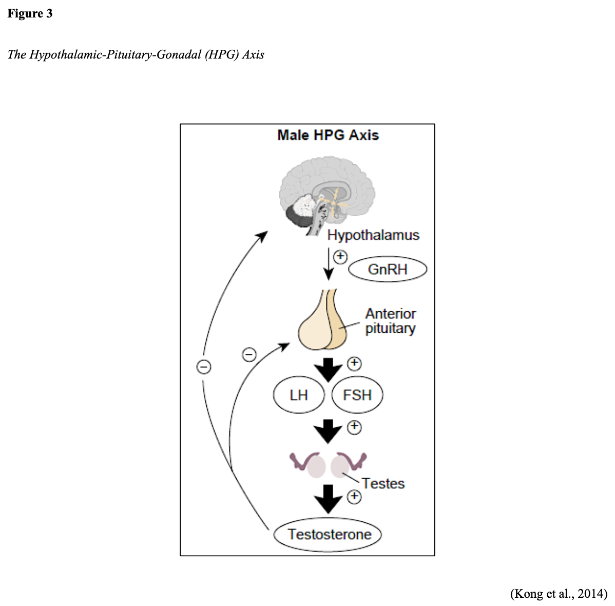
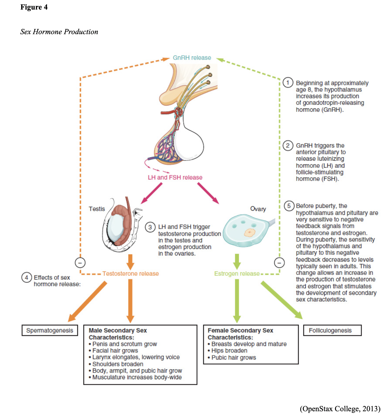
The production of androgens is initiated by the hypothalamus. The hypothalamus creates and releases gonadotropin-releasing hormone (GnRH), also called luteinizing hormone-releasing hormone (LHRH). GnRH prompts the pituitary gland to generate and secrete luteinizing hormone (LH) and follicle-stimulating hormone (FSH). Figure 4 compares this process in males and females. In males, testosterone production is regulated by luteinizing hormone (LH) and LHRH. LH acts on specific cells in the testes to produce testosterone; additional androgens are produced by the adrenal glands (Clavijo & Hsiao, 2018; Fujita & Nonomura, 2019).
Androgens are taken up by prostate cells, where they bind to the androgen receptor (AR) directly or are converted to DHT. DHT is found predominantly in prostate tissue and has a greater affinity for binding to the AR than testosterone. The AR is a steroid receptor transcriptional factor that mediates the actions of both testosterone and DHT. It has a similar structure and functionality to that of the estrogen receptor (ER) that fuels estrogen-based cancers, such as breast cancer in females. Androgens can fuel the growth of prostate cancer by binding to and activating the AR, which subsequently stimulates the overexpression of specific genes that trigger cell growth. Therefore, the AR serves a central role in prostate cancer, particularly castration-resistant prostate cancer (CRPC; Clavijo & Hsiao, 2018; Fujita & Nonomura, 2019).
Prostate Cancer Subtypes
All prostate cancers arise from glandular epithelial cells, which is the most abundant cell type within the prostate gland. More than 95% are adenocarcinomas, which are cancers that form in the mucus-producing glandular cells. Approximately 5% are transitional cell carcinomas, and less than 1% are squamous cell carcinomas. The thre
...purchase below to continue the course
Risk Factors
All men are at risk for prostate cancer; however, older age, African American race, and family history of prostate cancer are the most well-cited risks for the condition (US Preventative Services Task Force [USPSTF], 2018). As described earlier, African American men are at heightened risk. They tend to develop prostate cancer at a younger age and endure a more aggressive clinical course than other men. Family history is a significant risk factor, as up to 10% of all prostate cancers are hereditary. Men who have a first-degree relative with prostate cancer (i.e., father or brother) are twice as likely to develop prostate cancer as those with no family history. Men who have two or more first-degree relatives with prostate cancer are at a five-fold increased risk for prostate cancer (CDC, 2020). Other possible risk factors with weaker associations and less evidence include diets high in saturated fat and obesity; tobacco use is associated with a higher risk of mortality (USPSTF, 2018).
Genetic Mutations
Everyone has BRCA1 and BRCA2 genes. These genes function as tumor suppressor genes that work to prevent cancer by regulating specific cells’ growth and division under physiologic conditions. However, mutations in these genes prevent them from working correctly, thereby increasing cancer development’s propensity. Mutations in the BRCA1/BRCA2 genes are inherited in an autosomal dominant pattern. One copy of the mutated gene in each cell is sufficient to increase the risk of developing cancer. The altered gene can be inherited from either the male or female parent, as each child of a parent with a BRCA1 or BRCA2 mutation has a 50% chance of inheriting the same gene mutation. Most commonly known for increasing the risk of breast and ovarian cancers in women, mutations in the BRCA2 gene also increase the risk for aggressive prostate cancer in men. BRCA mutations occur most frequently within the Ashkenazi Jewish population. BRCA2 mutations are associated with earlier-onset, higher rates of lymph node involvement, distant metastasis at diagnosis, and higher mortality rates. Some studies describe the presence of a BRCA2 mutation as an independent prognostic factor for poorer outcomes in prostate cancer (Hartzfeld et al., 2016). Data from the IMPACT study (Page et al., 2019), which followed 2,900 men with annual PSA screenings for three years, demonstrated that BRCA2 mutation carriers had the most significant risk of developing cancer. Their findings revealed a 5.2% risk in BRCA2 mutation carriers, 3.4% risk in those with BRCA1 mutations, and a 2.7% risk in men who tested negative for both BRCA1/BRCA2 mutations (Page et al., 2019).
Genetic and Molecular Biomarker Analysis
Due to recent advancements in technology and targeted treatment modalities, the National Comprehensive Cancer Network (NCCN) guidelines recommend that tissue samples (currently only for patients with advanced or metastatic prostate cancer) undergo specialized genetic and molecular biomarker analysis. These specific analyses evaluate for the presence of homologous recombination gene mutations (HRRm) and microsatellite instability-high (MSI-H) or mismatch repair deficiency (dMMR). HRR gene mutations can increase the risk of developing more aggressive prostate cancer, and the most common include BRCA1/2, ATM, and CDK12. HRR gene mutations compromise tumor cells’ ability to correct the DNA damage through the loss of HRR. This mechanism can lead to an increased reliance on poly ADP-ribose polymerase (PARP)-mediated DNA repair pathways for survival, thereby opening up the possibility of targeted treatment pathways, such as the use of PARP-inhibitors (i.e., Olaparib [Lynparza]) in patients with advanced BRCA2-mutant prostate cancer. The presence of MSI-H or dMMR has clinical implications with regards to the potential use and benefit of immunotherapy (specifically, immune checkpoint inhibitors) in several types of cancers. MSI-H or dMMR are relatively uncommon in prostate cancer, with one study demonstrating a prevalence rate of only 3.2%. Their presence is still considered therapeutically meaningful in prostate cancer patients and is somatically acquired as the disease evolves. These mutations pose the potential for durable therapeutic responses to anti-programmed cell death protein 1 (PD-1)/programmed cell death ligand 1 (PD-L1) treatment, such as pembrolizumab (Keytruda), which will be discussed later within the treatment section of this module (Reichert et al., 2019).
Men with a genetic mutation in the HOXB13 gene may also be at increased risk for prostate cancer. HOX proteins are transcription factors that serve various roles in tissue differentiation of developing embryos, organogenesis, and contribute to several other cellular processes such as proliferation, differentiation, apoptosis, and migration (Brechka et al., 2017). The HOXB13 protein also acts as a tumor suppressor, which means that it keeps cells from growing and dividing too quickly or uncontrolled. While all individuals have two copies of the HOXB13 gene, mutations in one copy of the HOXB13 gene are associated with an increased risk for early-onset prostate cancer and CRPC. Research suggests that mutations in this gene impair the protein’s tumor suppressor function, resulting in uncontrolled cellular proliferation and cancer development (US National Library of Medicine [NLM], 2020). However, the clinical data is not nearly as robust as with BRCA2 mutations, and there remain numerous gaps in current understanding of how the HOXB13 gene confers increased risk in prostate cancer (Brechka et al., 2017).
Signs and Symptoms
Early prostate cancer generally does not cause pain, sexual dysfunction, or any other signs or symptoms. In most cases, affected men exhibit no symptoms. As the tumor grows or spreads into surrounding structures, patients may develop uncomfortable urinary symptoms, such as difficulty starting or stopping the flow of urine, a feeling of not being able to empty the bladder, or pain with ejaculation. However, many of these symptoms can also occur due to BPH, which is a common condition that affects a large percentage of men as they age. An enlarged prostate gland can induce uncomfortable urinary symptoms, including bladder, urinary tract, or kidney problems (Mayo Clinic, 2019). With advanced disease, symptoms are often the consequence of cancer metastasizing to other areas in the body. Based on cancer spread patterns, the most common symptoms include bone pain, dysuria, hematuria, flank pain, anemia, fatigue, weight loss, and weakness. (Leslie et al., 2020).
Early Detection and Screening
Given the lack of warning signs and symptoms in early prostate cancer, most men are diagnosed as a result of health screenings for a serum protein (prostate-specific antigen [PSA]) or from abnormalities detected on a digital rectal exam (DRE; Leslie et al., 2020).
PSA
PSA is a protein generated only by the prostate gland and a tumor marker for prostate cancer. Tumor markers are substances that may be produced by cancer or by the body’s response to cancer’s presence but are also made in smaller quantities by healthy cells, making them nonspecific. The PSA blood test measures the amount of this protein in the blood. A PSA level of less than 4.0 ng/mL was previously considered normal, and higher than 4.0 ng/mL was deemed abnormal, warranting a biopsy to evaluate for prostate cancer. Over time, these recommendations have changed. The PSA can be elevated in benign conditions such as BPH, prostatitis (infection or inflammation of the prostate gland), specific urologic procedures, or medications. Ejaculation, riding a bicycle, or strenuous exercise can induce a transient rise in PSA for a short time (Leslie et al., 2020; NCI, 2017). In contrast, commonly prescribed medications for BPH such as 5-alpha-reductase inhibitors or dihydrotestosterone blockers (i.e., finasteride [Proscar] and Dutasteride [Avodart]) are known to decrease PSA by approximately 50%, which can lead to false-negative results (NCCN, 2020). The American Board of Internal Medicine (ABIM) cites the PSA as having “no specific normal or abnormal level” in their 2020 Laboratory Test Reference Ranges (ABIM, 2020, p.9). In general, the PSA level varies over time and rises slowly with age, even in the absence of prostate abnormalities. Beyond a screening modality, the PSA test can help to stage, treat, and monitor prostate cancer (Cadet et al., 2019).
DRE
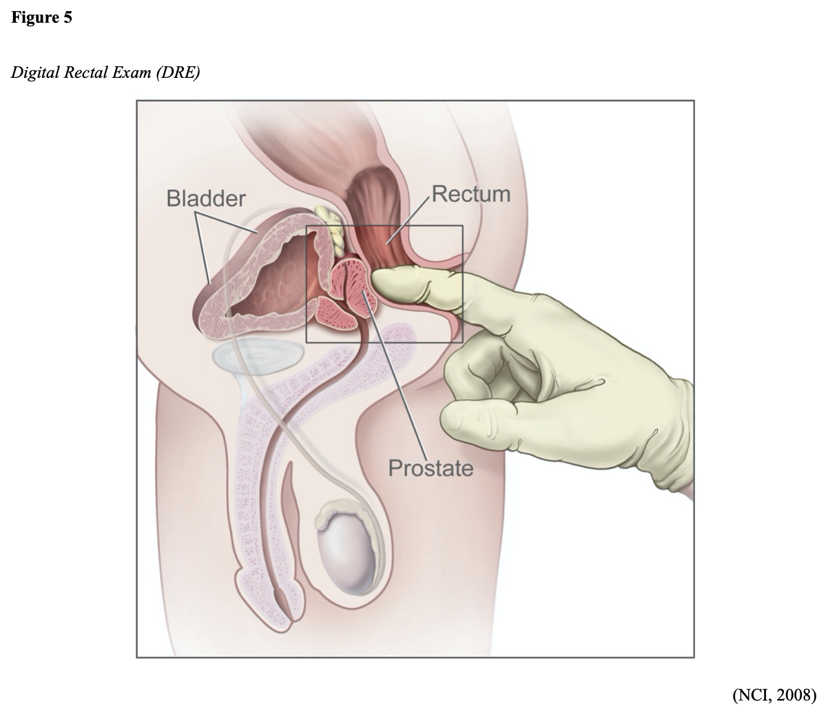
As demonstrated in Figure 5, the DRE allows for direct palpation of the PZ region, which is the site of most prostate cancers. The clinician performs this exam to feel for any bumps, nodularity, or abnormalities on the prostate gland that might suggest cancer (ACS, 2019b). Many clinicians, particularly primary care providers and urology specialists, regularly perform the DRE as part of their physical examination. However, research has demonstrated that the DRE carries a low specificity and sensitivity for detecting early prostate cancer. As of 2018, the US Preventive Services Task Force (USPSTF, 2018) stopped recommending DRE for early prostate cancer detection.
Controversies Over Prostate Cancer Screenings
Screening for prostate cancer is routinely performed throughout the US, but its benefit and value is controversial. It has been the subject of much criticism over the last few decades; it remains unclear if the benefits of prostate cancer screening outweigh the risks for most men. While prostate cancer screenings may lead to earlier diagnosis and treatment, this does not correlate with improved survival (Smith et al., 2019). The major shortcoming of prostate cancer screening is the lack of impact and questionable value of the information received, leading to disagreements about its importance amongst many experts. For men whose prostate cancer is detected by screening, it is impossible to predict who will benefit from treatment. Some men treated may avoid death and disability from prostate cancer, whereas others who are treated would have died of unrelated causes before their cancer became severe enough to spread, affect their health, or reduce the quality and longevity of life. PSA specificity and sensitivity are low, and screening is subject to high false-positive rates. A false positive entails an abnormal PSA level but no detectable prostate cancer, whereas a false negative is characterized by a normal PSA level despite the presence of cancer. Further, the PSA test has been criticized for flagging too many slow-growing cancers and subjecting patients to invasive procedures, interventions, treatments, and treatment side effects, when the cancer may never have posed a risk to the patient during their lifetime (Cadet et al., 2019; Leslie et al., 2020). The PSA level does not regularly correlate with CRPC, the most lethal type of prostate cancer (Hartzfeld et al., 2016), and there is an extensive literature base surrounding the overtreatment and overdiagnosis of prostate cancer due to PSA screenings (Kappen et al., 2019).
Prostate Cancer Screening Guidelines
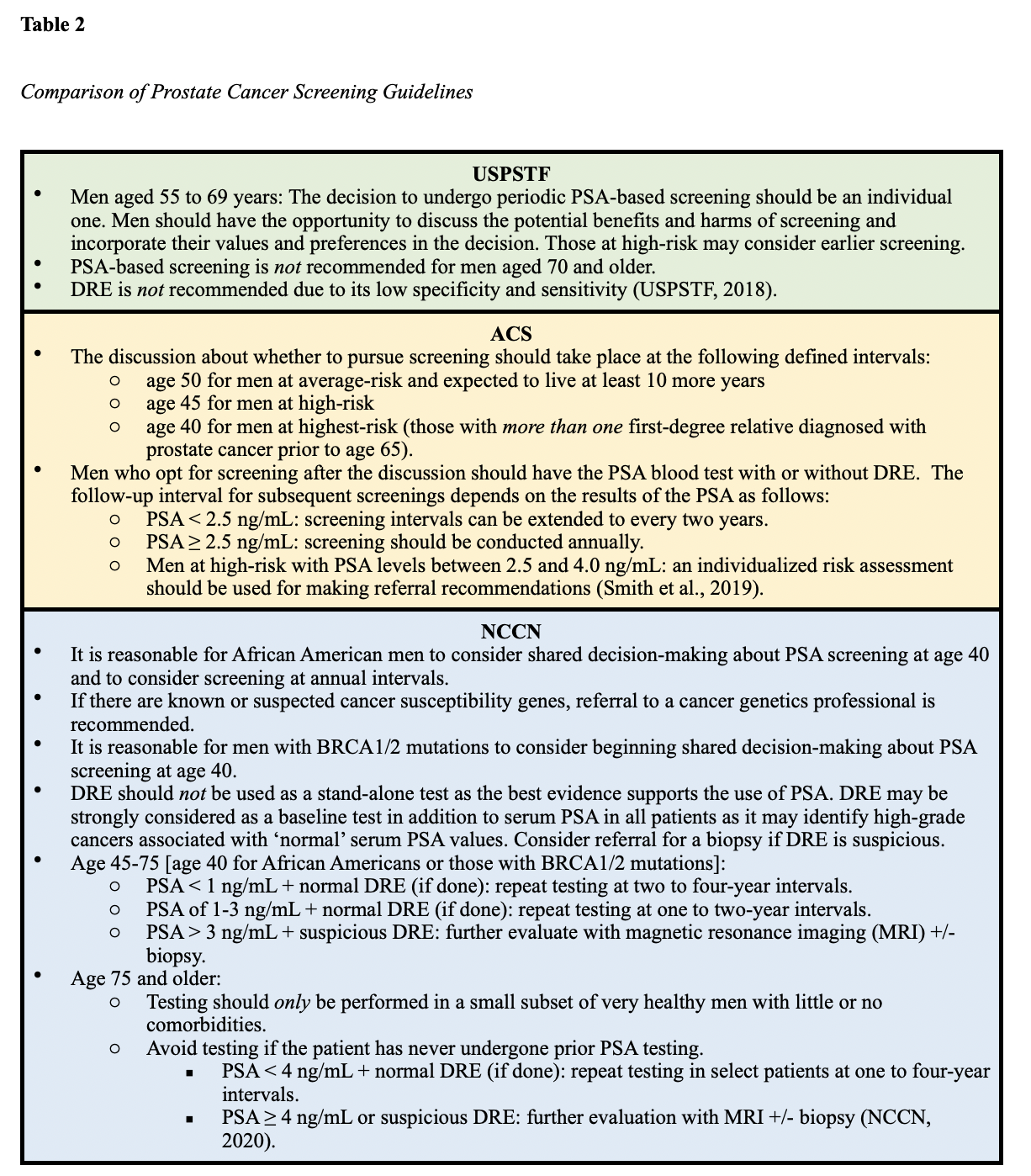
The current consensus from the USPSTF, NCCN, and ACS evidence-based screening guidelines is that the decision to perform PSA screening tests should be an individual one. Clinicians are encouraged to discuss the pros and cons of screening with patients and engage in shared decision-making before deciding upon screening. This offers men an opportunity to determine if screening is right for them and strives to reduce the overdiagnosis and overtreatment of prostate cancer. Given the controversies over prostate cancer screenings, there remain some discrepancies between the three guidelines. All three guidelines classify high-risk men as those who are African American or have a first-degree relative with prostate cancer. A summary of the specific screening guidelines according to each organization is outlined in Table 2 (ACS, 2019b; NCCN, 2020; USPSTF, 2018).
Based on the recent findings from the IMPACT trial discussed earlier, Page and colleagues (2019) recommend consideration of testing for BRCA mutations under the following circumstances:
- if there is a history of prostate, breast, or ovarian cancer in the immediate family, particularly among younger family members;
- if other family members test positive for BRCA1 or BRCA2 mutations;
- if the patient is of Ashkenazi Jewish descent (Page et al., 2019).
Diagnosis
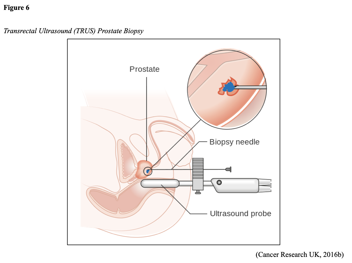
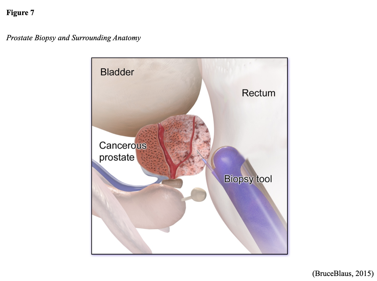
To ensure a thorough workup, the differential diagnosis for prostate cancer should always include BPH, prostatitis, and pyelonephritis, which share similar symptoms and may elevate PSA levels. Prostate cancer is classified by grade and stage, and staging can be clinical and pathological. Clinical staging is based on physical exam findings such as the DRE. While the PSA and DRE are useful in detecting the prostate gland abnormalities, tissue sampling is necessary to confirm prostate cancer and establish its clinical features. Pathological staging is more accurate and relies on the information acquired from surgery, such as the extent of the cancer invasion into surrounding structures. This allows for an enhanced understanding and accurate prediction regarding the patient’s treatment plan and prognosis (Cadet et al., 2019). A transrectal ultrasound-guided (TRUS) core biopsy of the prostate is the most common tissue sampling procedure. However, MRI or a ‘fusion’ of both imaging modalities is being used more frequently as technology advances. The needle is guided through the rectum (transrectal biopsy) or the skin between the scrotum and anus (transperineal biopsy) to remove a small prostate tissue core. This process is repeated several times to obtain up to 12 core tissue samples. The TRUS procedure is demonstrated in Figures 6. Figure 7 provides a magnified view of a prostate biopsy regarding the anatomical location of the prostate (ACS, 2017b; Leslie et al., 2020).
Gleason Score
All prostate cancers are subjected to a histologic grading system called the Gleason score, which is vital in predicting prostate cancer’s behavior and determining the best treatment options. Initially developed in the 1960s, the Gleason score is the most widely used and reliable system to define prostate cancer’s aggressiveness (Leslie et al., 2020). The Gleason score is based on the cellular architecture, microscopic arrangement, and pattern of the glands within the prostate tissue. This is distinct from other cancers, which are generally defined by individual cellular characteristics. To determine the Gleason score, a histologic grade ranging from one to five is assigned to the tissue. This represents how abnormal the cancer cells look under a microscope and how aggressive the cancer appears. As demonstrated in Figure 8, if the cells appear very similar to normal, healthy prostate tissue, they are considered well-differentiated, and a grade one is assigned. If the cells are markedly abnormal and do not have any characteristic features of healthy cells, they are poorly-differentiated, and a grade five is assigned. Grades two through four have features between these extremes. Low-grade cancer cells tend to grow and spread more slowly than high-grade cancer cells. Two grades are assigned; one for the most predominant (primary) pattern identified within the tissue sample, and one for the second most predominant pattern. Next, the two grades are added together to form the Gleason score, which ranges between 6 and 10 in modern-day clinical practice (Leslie et al., 2020; Prostate Cancer Foundation [PCF], n.d.).

In 2014, the International Society of Urological Pathology (ISUP) developed a revised prostate cancer grading system called the Grade Groups. The system is more straightforward and was devised to enable patients to understand their actual risk level better and limit overtreatment. The new system compresses the Gleason score into grade groups, ranging from one to five, as represented in Table 3. For example, if the Gleason score is presented as 3+4=7, this means that the tumor is primarily graded three, and less of the tumor is a grade four. When combined, they are a Gleason score of 7. If a tumor is all the same grade (i.e., grade 4), then the Gleason score is reported as 4+4=8 (ACS, 2017). The new system has since been adopted by the World Health Organization (WHO) and incorporated in the NCCN evidence-based guidelines. Compiled through rigorous clinical trial research and annual expert panel reviews, the NCCN provides evidence-based treatment guidelines for each cancer type according to distinct features such as cellular characteristics, genetics, staging, and inheritance patterns. The guidelines are widely utilized by the vast majority of NCI-accredited cancer centers and help to guide medical decision making throughout the patient’s disease trajectory (NCCN, 2020).

Prostate Cancer Staging
Prostate cancer is grouped into four stages, which are defined by the extent of the tumor presence and how rapidly the cancerous cells are growing. The four stages are determined by the Gleason Score and the Tumor, Nodes, and Metastasis (TNM) system. In the TNM system, T stands for the primary tumor and is a measure of the tumor’s size and extent, N stands for lymph node(s) and is based on whether cancer has spread into nearby lymph nodes, and M stands for metastasis and based on whether cancer has spread to distant sites in the body. The T is labeled on a scale of TX through T4, where TX designates that the primary tumor cannot be assessed, and T0 refers to no evidence of the primary tumor. T1 and T2 denote early prostate cancer, and T3 and T4 indicate advanced prostate cancer (NCCN, 2020; Urology Care Foundation, 2019). Table 4 depicts the fundamental distinctions in the clinical and pathological staging of prostate cancer based on the TNM system, as extracted from the 8th edition of the American Joint Committee on Cancer [AJCC] Staging Manual (2018).
Table 4
TNM Staging System

Principles of Imaging Studies
The only way to definitively determine the presence or absence of distant metastases is through radiographic imaging. However, unlike most cancer diagnoses, systemic imaging studies are not universally recommended for all patients diagnosed with biopsy-proven prostate cancer. According to the NCCN (2020), the goal of prostate cancer imaging is to detect and characterize disease to develop the proper treatment regimen and guide a change in management. The NCCN guidelines offer specific recommendations regarding which men require imaging studies as part of their initial staging workup.
- Imaging is not indicated for men who meet all of the following criteria:
- T1-T2a,
- grade Group 1,
- PSA < 10 ng/mL, and
- fewer than three prostate biopsy cores positive, and ≤50% cancer in each core.
- Men who have any features outside the above should undergo the following imaging studies as part of their initial staging workup:
- bone imaging with a radionuclide bone scan or single-photon emission computerized tomography (SPECT) imaging. A bone scan is also indicated in the initial evaluation of patients at high-risk for bone metastases and any patient with symptoms consistent with bone metastases.
- imaging to evaluate the anatomic or functional parameters of the pelvic region and abdomen.
- Anatomic imaging techniques may include plain film radiographs (x-rays), ultrasound, computerized tomography (CT scans), or standard MRI.
- Functional imaging techniques can consist of positron emission tomography/computed tomography (PET/CT) scan, sodium fluoride PET/CT scan, or advanced MRI techniques such as multi-parametric magnetic resonance imaging (mpMRI). A mpMRI is preferred over standard MRI imaging, as this test can generate more detailed pictures of the prostate (NCCN, 2020).
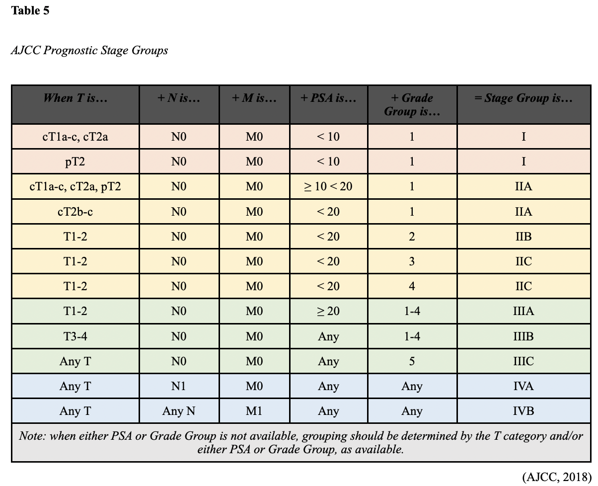
The information acquired from above is subsequently applied to the algorithm represented in Table 5 to determine the definitive stage of prostate cancer.
Management
Watchful Waiting
Not all men diagnosed with prostate cancer will require treatment, as it is characteristically slow-growing, and in many cases, patients die from other causes. Active surveillance or “watchful waiting,” is usually a reasonable option for early-stage prostate cancer patients. The NCI dictionary of cancer terminology defines watchful waiting as:
“Closely watching a patient’s condition but not giving treatment unless symptoms appear or change. Watchful waiting is sometimes used in conditions that progress slowly. It is also used when the risks of treatment are greater than the possible benefits. During watchful waiting, patients may be given certain tests and exams. Watchful waiting is sometimes used in prostate cancer. It is a type of expectant management” (NCI, n.d.).
In clinical practice, watchful waiting can provide valuable information regarding how quickly the prostate cancer is progressing, if at all. It typically involves long-term monitoring with modalities such as periodic PSA testing, DRE exams, prostate biopsies, symptom assessment, and imaging studies until clinicians and patients decide that treatment should be implemented. Most surveillance protocols recommend PSA monitoring every three to six months and a repeat biopsy after the first year, and as clinically indicated. Watchful waiting can induce significant anxiety for patients and their families, who may be apprehensive about not treating cancer. It is crucial that APRNs counsel patients and families on the rationale for this approach and clarify that watchful waiting is not a euphemism for ‘doing nothing.’ Ultimately, the goal is to delay treatment in favor of careful monitoring for cancer progression to preserve the patient’s s quality of life and reduce the risk of complications and side effects of treatment. According to the NCCN (2020), the standard of care most commonly used when recommending watchful waiting for early-stage prostate cancer is a ten-year life expectancy. If a patient is reasonably expected to benefit from more aggressive treatment for ten years or more, the recommendation is usually to pursue treatment instead (NCCN, 2020; Urology Care Foundation, 2019).
Treatment Modalities
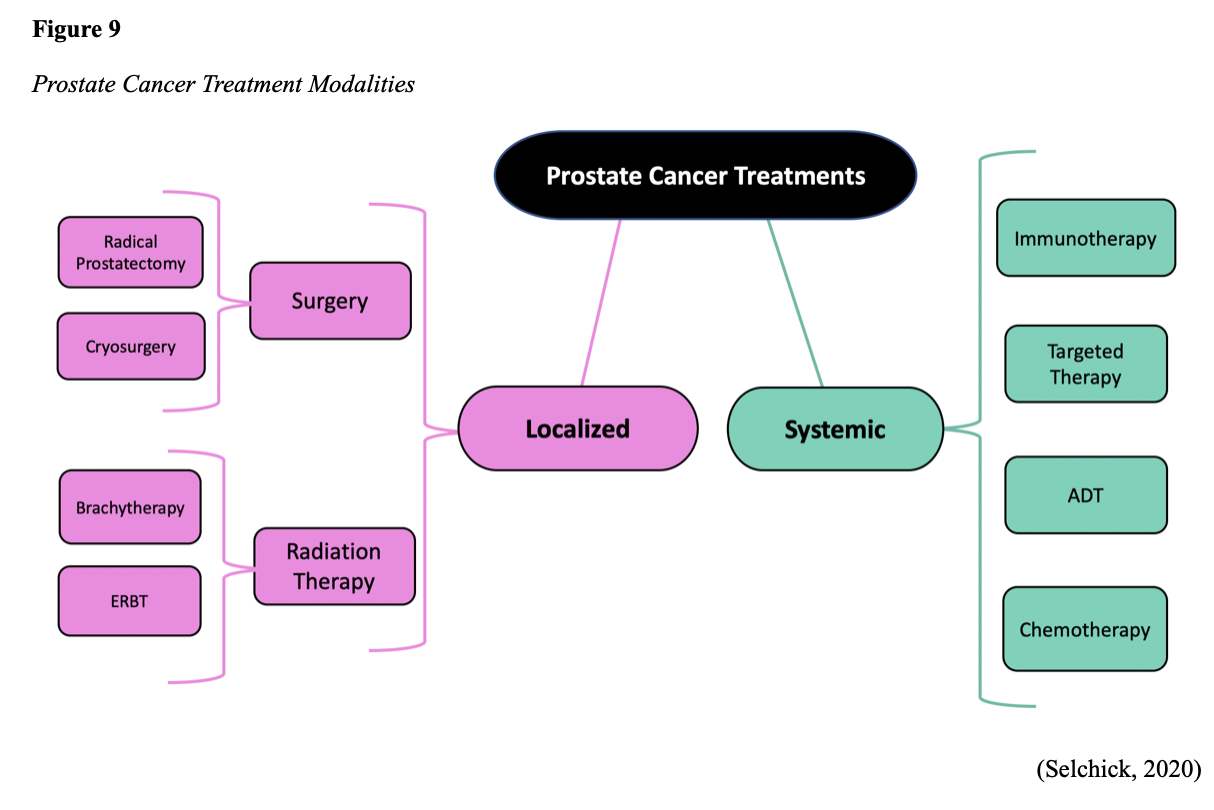
The decision to initiate treatment is multifactorial and based on the patient’s age, life expectancy, coexisting medical conditions, specific genetic mutations, symptom burden, and cancer stage. Treatment for prostate cancer is divided into two major categories; localized and systemic therapies, as outlined in Figure 9. Patients with early-stage and localized disease are treated with curative intent. These patients are most commonly offered surgical intervention, followed by adjuvant (post-surgery) radiation therapy. Patients with advanced or metastatic prostate cancer are more likely to be treated with a combination of systemic therapies and are subject to relapse and remission (NCCN, 2020).
Radical Prostatectomy
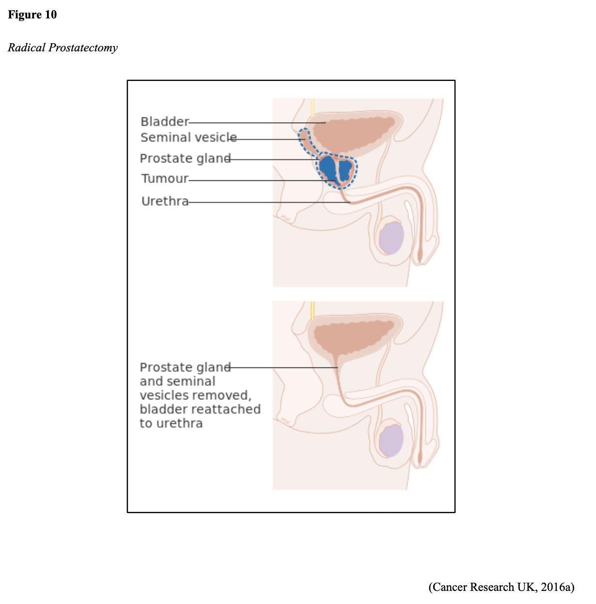
If the cancer is confined to the prostate, then a radical prostatectomy is the most common procedure. As demonstrated in Figure 10, the entire prostate gland, and usually the seminal vesicles, surrounding tissue, lymph nodes, nerves, and veins are removed. A portion of the urethra is also removed, and the remaining urethra is anastomosed at the bladder neck. A urinary catheter is temporarily inserted to drain the urine and allow the patient to heal (ACS, 2019c).
Erectile dysfunction (ED) or impotence is one of the most common risks associated with radical prostatectomy. Since the nerves which control a man’s ability to have an erection are within proximity to the prostate gland, they may become damaged, severed, or removed during the surgery. Approximately 50% of men with intact nerves will regain some ability to have an erection, but it can take six to twelve months. Men with other health conditions that impair their ability to maintain an erection, such as diabetes or vascular issues, will have a more difficult time regaining their normal function (University of Michigan Health System [UMHS], 2019). To mitigate the risk and burden of ED, many urologists have begun to implement the relatively novel nerve-sparing radical prostatectomy technique; however, ED continues to be problematic for many patients postoperatively (JMU, n.d.). Urinary incontinence and damage to the urethra is another common risk associated with radical prostatectomy. Once the urinary catheter is removed postoperatively, up to 50% of men will develop urinary incontinence. The degree of incontinence can range from mild and intermittent dribbling, stress incontinence, or continuous leakage requiring the use of pads or incontinence briefs. If the incontinence persists for longer than one year, some men may need additional urologic surgery (UMHS, 2019).
Cryotherapy
Cryotherapy, also referred to as cryosurgery or cryoablation, is a minimally invasive surgery that uses thin needles and controlled freezing gas to destroy the cancer cells. It is a relatively novel and revolutionary treatment modality but still lacks rigorous, long-term survival data. Currently, it is considered a treatment option for men with localized or locally advanced prostate cancer. According to the NCCN (2020) guidelines, cryotherapy is reserved for PSA persistence or recurrence after radical prostatectomy. PSA persistence/recurrence is defined by the NCCN (2020) as follows: “the failure of PSA to fall to undetectable levels (PSA persistence) or undetectable PSA after radical prostatectomy with a subsequent detectable PSA that increases on two or more determinations (PSA recurrence)” (NCCN, 2020, p. PROS-11). Cryotherapy is also an option for radioresistant disease, which refers to prostate cancer that does not respond to radiation or recurs after radiation therapy. Prostate cryotherapy may be used alongside other treatments for prostate cancer. Since cryotherapy destroys both healthy and cancerous tissue that it comes in contact with, ultrasound guidance is used to facilitate the placement of the cryoprobes into the prostate. Argon gas is subsequently injected into the prostate, promoting the formation of ice crystals inside and around the cells. The freezing and thawing process destroys the cancer cells by dehydrating them, causing extreme changes in the pH levels, and preventing blood flow, blocking necessary nutrients. Some sources cite that by subjecting the prostate gland to freezing gas of -40° C, an anti-tumor response is activated within the body, and antibodies are produced to eradicate the tumor. The procedure is generally well-tolerated, but most patients experience soreness and hematuria for one or two days following the procedure. Potential complications include urinary incontinence, injury to the rectum, and erectile dysfunction. The side effects tend to worsen in patients who have had prior radiation therapy (ZERO - The End of Prostate Cancer, n.d.).
Radiation Therapy
Up to 50% of men have pathologic extension beyond the prostate gland at radical prostatectomy. Therefore, the role of adjuvant radiation therapy has evolved over the last few decades. The NCCN (2020) supports the use of radiation therapy for adverse laboratory or pathologic features following surgery, which include positive surgical margin(s), seminal vesicle invasion, extracapsular extension, or detectable PSA (NCCN). Radiation therapy may also be used as the first-line treatment for early-stage and low-grade prostate cancer confined to the prostate gland, or as part of the treatment regimen alongside hormonal therapy. Curative rates for men in this category are nearly equivalent to those who undergo radical prostatectomy. Across clinical studies, research consistently demonstrates that both treatment modalities are associated with satisfactory clinical outcomes in terms of disease control in localized prostate cancer (ACS, 2019a; Tamada et al., 2017). Beesley and colleagues (2019) conducted a cohort study, including 4,544 patients, and found no apparent difference in clinical outcomes between surgery and radiotherapy on average.
Radiation therapy has dramatically evolved since the original use of Cobalt therapy machines in the 1950s. Scientific advancements have led to the development of computers for treatment planning and dose optimization, which help minimize treatment-related toxicities (Yarbro et al., 2018). Radiation therapy is a type of localized cancer treatment that uses high-energy rays or electron, proton, or neutron beams to kill cancer cells. The primary objective is to deliver a precisely measured dose of radiation to a defined tumor with as little injury as possible surrounding healthy tissue. Radiation induces cellular damage to cancer cells, leading to biological changes in the DNA. This causes cells to die over days, weeks, and months, rendering them incapable of reproducing or spreading. All healthy cells and cancer cells are vulnerable to the effects of radiation and may be injured or destroyed; however, most normal cells can repair themselves and remain functional. The total dose of radiation is hyper-fractionated, which means it is administered in smaller divided doses, or fractions, rather than all at once. Hyper-fractionation allows healthy cells a chance to recover between dosing. Each dose is called a fraction, and the total number of fractions depends on the tumor size, location, reason for treatment, patient’s overall health, performance status, goals of therapy, as well as consideration of any other treatments the patient is receiving. Radiation can be delivered externally or internally, and some patients may receive both types (Yarbro et al., 2018).
Currently, the two most common types of radiotherapy used for prostate cancer include internal brachytherapy and external beam radiation therapy (EBRT) and will be described in this subsection (NCCN, 2020). However, radiation is an evolving subsector of cancer medicine, and it is essential to acknowledge that several additional advanced radiotherapy techniques are increasingly applied to this population. As scientific advancements and clinical data evolve within this rapidly growing cancer care field, some of the novel radiation treatments listed below may eventually replace our preexisting treatment modalities:
- three-dimensional conformal radiotherapy (3D-CRT),
- image-guided radiation therapy (IGRT),
- intra-operative radiation therapy (IORT),
- proton beam radiation therapy (PBRT),
- tomotherapy,
- hypofractionated radiation therapy,
- systemic radiation therapy,
- radioimmunotherapy (RIT; ACS, 2019a).
Brachytherapy
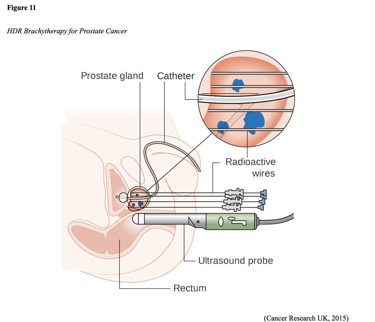
Prostate brachytherapy is a minimally invasive radiation therapy that implants a small wire or radioactive seeds directly into the prostatic tissue. Brachytherapy delivers high and concentrated doses of radiation to the prostate gland from inside the body. Hormone therapy is sometimes given for three to six months before brachytherapy to shrink the prostate gland and optimize the brachytherapy effectiveness. The two brachytherapy types include a temporary high-dose-rate (HDR) or a permanent low-dose-rate (LDR). HDR brachytherapy is demonstrated in Figure 11 and is usually performed on an outpatient basis under general anesthesia. Several catheters, which are attached to a machine that contains radioactive pellets, are placed into the prostate gland. The catheters transmit concentrated bursts of high-dose radiation pellets into the tumor bed. This is generally performed once or twice and takes about 15-minutes (Memorial Sloan Kettering Cancer Center [MSKCC], n.d.).
LDR brachytherapy also requires general anesthesia and is generally performed on an outpatient basis. It involves placing permanent radioactive seeds into the prostate gland, slowly releasing radiation over several months. Up to 100 seeds may be inserted depending on the size of the prostate. The radiation emitted from the seeds only travels a short distance, limiting the amount of damage to nearby healthy tissues. The radioactivity lessens after several weeks or a few months. Patients are usually advised to stay at least three feet away from pregnant women and small children for a specified period. This is recommended to ensure any unnecessary risk is avoided. While there is little clinical data available to validate this recommendation and the duration of time is not yet defined, the statement is endorsed by the International Atomic Energy Agency (IAEA, n.d.) within their Radiation Protection of Patients (RPOP) recommendations. Patients should be counseled that detection systems can sometimes pick up low radiation levels at airports, so they should carry a physician’s note or radiation card regarding their treatment to avoid security problems. Overall, patients can safely go about their regular routines and lifestyles without potentially exposing others (ACS, 2019a; MSKCC, n.d.).
EBRT
EBRT is radiation delivered from a source outside the body, directly to the cancer site. ERBT uses a linear accelerator to generate and deliver high-energy x-rays in small daily doses. Intensity-modulated radiation therapy (IMRT) is the most common type of EBRT for prostate cancer. It uses a computerized machine that travels around the patient as it delivers the radiotherapy. This technology allows for the more precise shaping and aiming of the radiation beams at the prostate, covering several angles and allowing for increased intensity (strength) of the beams. This limits the dose of radiation to the surrounding healthy tissues and enables the patient to tolerate a higher radiation dose to cancer, improving clinical outcomes and survival (ACS, 2019a; Fischer-Valuck et al., 2018).
Radiation Side Effects. According to JMU (n.d.), about 25 to 50% of men who undergo brachytherapy will experience ED following prostate brachytherapy compared to nearly 50% of men who undergo EBRT. LDR brachytherapy also carries a risk for the migration of the implanted seeds. Additional side effects of radiation include fatigue, lymphedema in the legs or genital region, urinary incontinence, cystitis (inflammation of the bladder), urethral stricture, and proctitis (inflammation of the rectum; ACS, 2019a).
For a more in-depth review of radiation therapy and nursing implications, refer to the Oncology Nursing Part 1: Surgical and Radiation Oncology NursingCE course and earn 5 ANCC contact hours.
Androgen Deprivation Treatment (ADT)
ADT is also commonly referred to as testosterone-depleting therapy. Since testosterone cannot discriminate between healthy tissue receptors and those of cancerous tissue, ADT is the most common systemic treatment modality for prostate cancer. It deprives the body of androgens by lowering (or depleting) the testosterone level, which fuels cancer growth. Prostate cancer usually stops growing in response to hormone therapy, at least for a while. CRPC occurs when cancer outsmarts or becomes resistant to ADT therapy and begins to grow again despite the hormone-blocking treatments. The most common approaches to androgen deprivation include castration, antiandrogens, and combined-androgen blockade. A castrate level refers to having 90 to 95% less testosterone than a healthy male, and castration may be achieved surgically or chemically. Orchiectomy is the surgical removal of the testicles, a permanent and irreversible procedure that can reduce the testosterone level in the blood by 90 to 95%. Chemical castration is much more common and involves the use of LHRH analogs or LHRH antagonists (NCI, 2019).
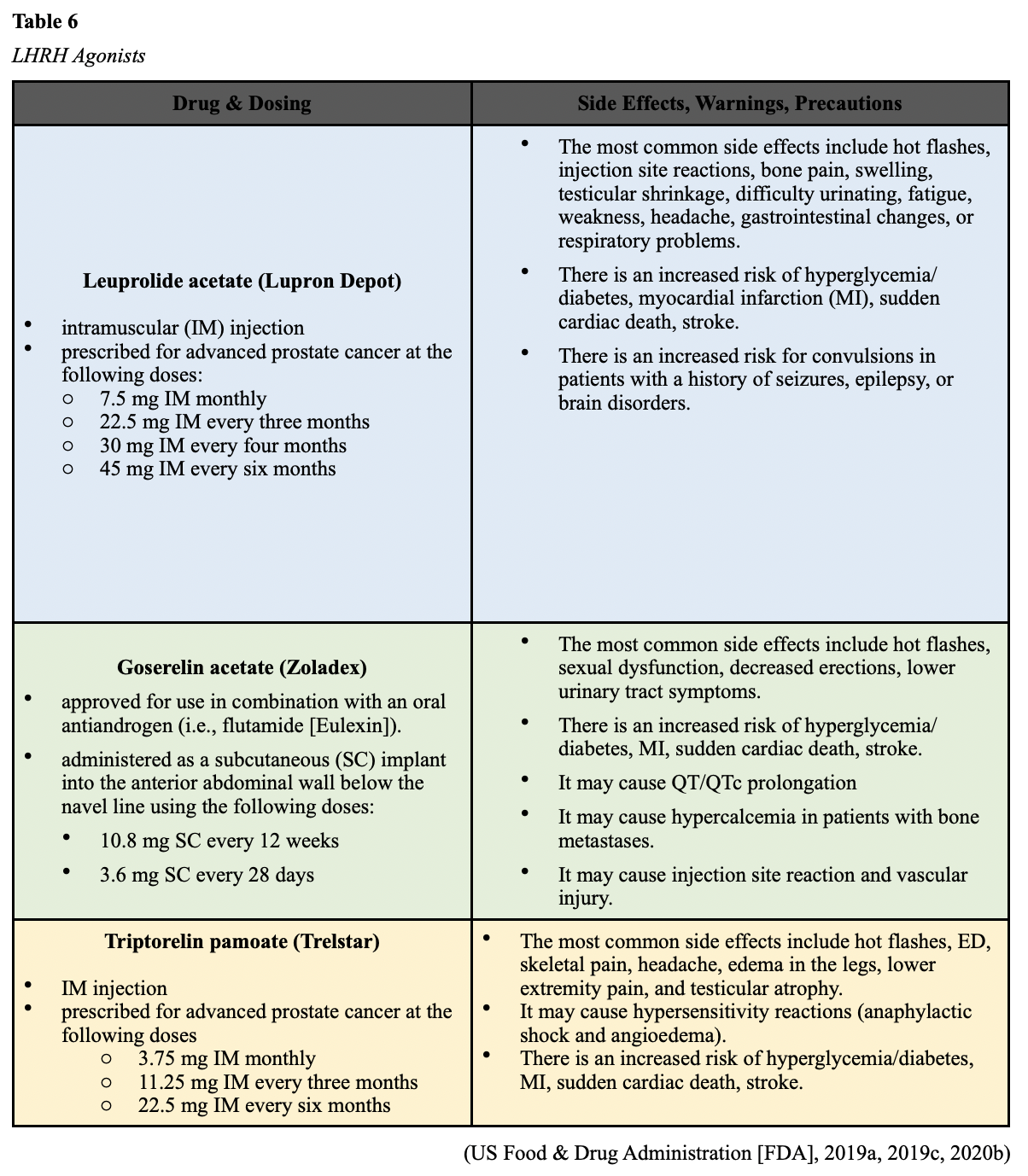
LHRH Agonists
LHRH agonists (sometimes called LHRH analogs) mimic LHRH’s action by occupying the receptors’ pituitary glands. Administration of these drugs causes the testicles to temporarily increase testosterone production, igniting a “testosterone flare” before blocking its release. Since the LHRH agonist has a longer half-life than physiologic LHRH, the LHRH agonist occupies the AR with greater affinity. Over time, the continued presence of high levels of LHRH agonists triggers the pituitary gland to stop producing LH, thereby halting the production of testosterone and reducing the body’s testosterone to castrate level. The testosterone flare is usually counteracted by the concurrent use of oral antiandrogen therapy (described below) for several treatment weeks. LHRH agonists are administered by injection or may be implanted under the skin. Currently, the three primary LHRH agonists used to treat prostate cancer in the US include leuprolide acetate (Lupron Depot), goserelin acetate (Zoladex), and triptorelin pamoate (Trelstar). These agents are outlined below in Table 6. All medications in this class are associated with an initial testosterone flare, which may cause an acute worsening of symptoms. This is particularly concerning for patients with advanced prostate cancer as it can lead to significant bone pain and ureter or bladder outlet obstruction (NCI, 2019). It can induce spinal cord compression (SCC) in patients with metastatic disease to the spinal column in severe cases. SCC is the compression of the spinal cord by malignant tumor invasion into the epidural space, and symptoms may include the following:
- sudden onset of acute back pain that worsens in the supine recumbent position and may be relieved by sitting,
- neurologic dysfunction such as numbness,
- bowel or bladder dysfunction such as incontinence, passing very little urine, or inability to urinate or defecate,
- altered gait or difficulty ambulating.
SCC is an oncologic emergency that requires immediate attention to preserve function and reduce morbidity. Initial treatment is critical and involves administering corticosteroids to reduce vasogenic edema in the spinal cord, thereby improving neurologic dysfunction and relieving pain, followed by definitive treatment with either radiation therapy or surgery (Klemencic & Perkins, 2019).
LHRH Antagonists
LHRH antagonists may also be called GnRH antagonists. These agents work by preventing LHRH from binding to its receptors in the pituitary gland, thereby inhibiting LH and testosterone production. Unlike LHRH agonists, LHRH antagonists do not cause a testosterone flare. Degarelix (Firmagon) is currently the only LHRH antagonist approved for prostate cancer treatment in the US (NCI, 2019). Degarelix (Firmagon) is a subcutaneous injection, and treatment is initiated at a dose of 240 mg, followed by a maintenance dose of 80 mg every 28 days. The most common side effects include injection site reactions (localized pain, erythema, swelling or induration), hot flashes, and weight gain. Hepatic laboratory abnormalities are also reported, particularly increased transaminases and gamma-glutamyl transferase (GGT) levels. Degarelix (Firmagon) carries a risk for QT/QTc interval prolongation, requiring periodic cardiac monitoring, and hypersensitivity reactions, which manifest as anaphylaxis, urticaria, and angioedema (FDA, 2015a).
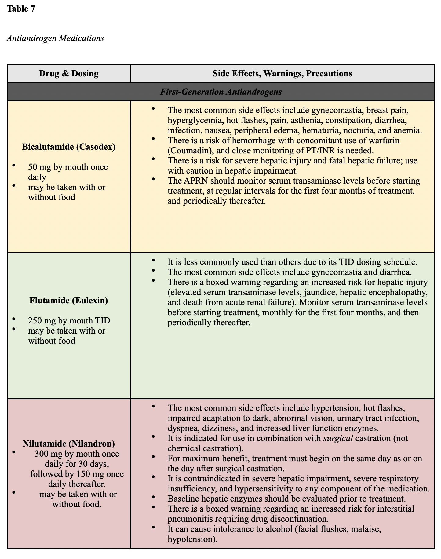
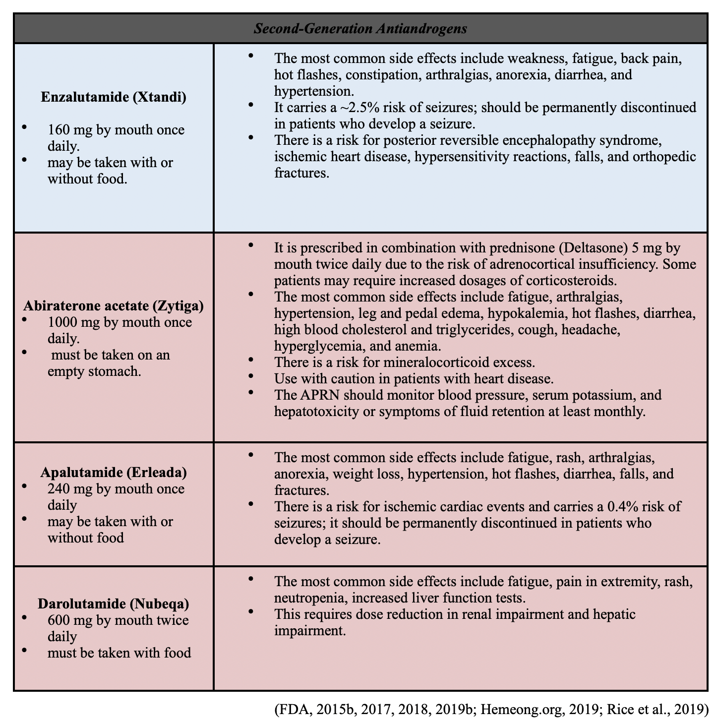
Antiandrogen Therapy
Antiandrogens are also called androgen receptor inhibitors or androgen receptor antagonists. These medications differ from LHRH antagonists by blocking particular facets of androgen signaling. They compete with androgens by binding to the AR. Testosterone circulates through the body but cannot interact with the prostate gland or promote cancer growth. Antiandrogens are oral medications that are rarely used as monotherapy since they do not block testosterone production. Instead, they are prescribed concurrently with LHRH agonists or in patients who have undergone a bilateral orchiectomy. This combined regimen is referred to as a combined androgen blockade. There are two major classes of antiandrogens; first-generation and second-generation. First-generation antiandrogens are credited with establishing the androgen receptor blockade as an effective treatment strategy in prostate cancer; however, they do not completely block all androgen receptor activity. These agents primarily block androgen production in the testes. First-generation antiandrogens currently approved for use in the US include bicalutamide (Casodex), flutamide (Eulexin), and nilutamide (Nilandron). Second-generation antiandrogens were developed to improve upon the mechanisms of first-generation drugs and in an attempt to bypass resistance to therapy. Second-generation agents have increased specificity and higher affinity for androgen receptors. Abiraterone acetate (Zytiga) was the first second-generation antiandrogen approved by the FDA in 2011. It is slightly different than others in the class as it works by preventing androgen biosynthesis and testosterone production in the testes, the adrenal glands, and the tumor. There are four second-generation antiandrogens currently approved by the FDA: enzalutamide (Xtandi), abiraterone acetate (Zytiga), apalutamide (Erleada), and darolutamide (Nubeqa). These agents are described in Table 7 (NCI, 2019; Rice et al., 2019).
Chemotherapy
Chemotherapy works by destroying quickly dividing cells and is not commonly used as an upfront treatment option for prostate cancer. Due to the slow-growing nature of the disease and the high toxicity associated with chemotherapy, chemotherapy is generally reserved for more advanced, widespread disease stages that have become refractory to the effects of ADT and antiandrogen therapy. When used as salvage therapy for progressive disease, chemotherapy has shown success in extending the longevity and quality of life in many patients. As a class, chemotherapy encompasses a group of high-risk, hazardous drugs that are administered with the intent to destroy as many cancer cells with as minimal effect on healthy cells as possible. Premised on cellular kinetics concepts, chemotherapy generally works by interfering with the normal cell cycle, impairing DNA synthesis and cell replication, which thereby prevents cancer cells from dividing, multiplying, and forming into new cancer cells. Since cancer cells tend to divide rapidly, chemotherapy targets cells that divide quickly. As a result, it also impacts healthy cells that divide quickly, such as those within the gastrointestinal tract, skin/hair cells, and bone marrow, which is why the most common chemotherapy side effects include bone marrow suppression, nausea, vomiting, diarrhea, fatigue, hair loss, and mucositis (Yarbro et al., 2018). The most common chemotherapy agents used for prostate cancer include docetaxel (Taxotere), paclitaxel (Taxol), Cabazitaxel (Jevtana), carboplatin (Paraplatin), and vinblastine (Velban). The side effects, dosing, and administration guidelines vary based on the agent and the patient’s clinical status. Less commonly, an agent called mitoxantrone (Novantrone) may be used but is only recommended for palliation in symptomatic patients who cannot tolerate other therapies (NCCN, 2020).
For a more in-depth review of radiation therapy and nursing implications, refer to the Oncology Nursing Part 2: Chemotherapy and Oncologic Emergencies and Oncology Administration NursingCE courses and earn up to 12 ANCC contact hours.
PARP Inhibitors
PARP inhibitors are a class of targeted agents referred to as small molecule inhibitors that block the PARP. This protein has a critical role in cell growth, regulation, and repair, helping cancer cells repair themselves and survive. This inhibition leads to cancer cell death. PARP-inhibitors have revolutionized how BRCA–mutant cancers, such as ovarian cancer and breast cancers, are treated. Their value has recently been discovered in BRCA-mutant prostate cancers (Yarbro et al., 2018). On May 15, 2020, the FDA granted accelerated approval of rucaparib (Rubraca) for patients with BRCA-mutant, metastatic CRPC who have been previously treated with androgen receptor-directed therapy and taxane-based (i.e., docetaxel [Taxotere] or paclitaxel [Taxol]) chemotherapy. Rucaparib (Rubraca) is dosed at 600 mg orally twice daily with or without food. Patients receiving rucaparib should also receive an LHRH agonist concurrently or should have had previously undergone bilateral orchiectomy. The most common side effects include fatigue, anemia, anorexia, rash, nausea, vomiting, diarrhea, constipation, thrombocytopenia, and increased transaminase levels (Oncology Nursing Society [ONS] Voice, 2020). Olaparib (Lynparza) is another oral PARP-inhibitor that has proven efficacy in treating BRCA-mutant breast and ovarian cancers. Olaparib (Lynparza) is currently being studied with regard to its clinical usefulness for BRCA-mutant prostate cancers. Recent clinical results from the Targeted Agent and Profiling Utilization Registry (TAPUR) Study were presented at the 2020 American Society of Clinical Oncology (ASCO) Annual Meeting. In two small cohorts, olaparib (Lynparza) resulted in objective responses or stable disease for at least 16 weeks in more than two-thirds (68%) of patients with BRCA1 or BRCA2-mutant advanced prostate cancer (ASCO, 2020).
Immunotherapy
Sipuleucel-T (Provenge) is a cellular immunotherapy customized using each patient’s immune cells to create a vaccine. The patient’s immune cells are collected through a process called leukapheresis and then developed into a vaccine devised to boost the patient’s immune system’s ability to attack prostate cancer. The cells are subsequently returned to the patient through an IV infusion about 72 hours later. The patient is given three doses, about two weeks apart. Sipuleucel-T (Provenge) is only currently approved for use in patients with metastatic CRPC who are asymptomatic or minimally symptomatic. Further, eligible patients must not have liver metastases and have a life expectancy greater than six months to receive this medication. In phase 3 clinical trial, Sipuleucel-T has been shown to extend average survival by only about four months; however, this translates to a 22% reduction in mortality risk for patients in this category. The infusion is generally well-tolerated, and the most common side effects include fever, chills, infusion-related reactions, and headache (NCCN, 2020).
Pembrolizumab (Keytruda) is a type of immunotherapy that blocks the PD-1 pathway and triggers the immune system to recognize the cancer cells as foreign and attack them. Pembrolizumab (Keytruda) has demonstrated clinically significant results, including improved survival and quality of life across various cancer types. With regards to prostate cancer, pembrolizumab (Keytruda) is currently only approved for use as subsequent systemic therapy for patients with MSI-H or dMMR mutation who have progressed through at least one line of systemic treatment for metastatic CRPC. As cited earlier, MSI-H and dMMR mutations are relatively uncommon in prostate cancers; however, the pembrolizumab (Keytruda) has demonstrated remarkable and durable responses in those who express these mutations. Abida and colleagues (2018) found that in 11 patients with MSI-H/dMMR CRPC, six patients (54.5%) had more than a 50% decline in PSA levels, while four patients had radiographic responses with reduction of tumor burden on imaging. Further, five of the six responders remained on treatment, with the most prolonged ongoing response extending 89 weeks. While the number of patients with MSI-H/dMMR CRPC is small and clinical research continues, these findings support the clinical significance of this mutation with regards to treatment planning (Reichert et al., 2019).
Prevention of Skeletal-Related Events (SREs)
Since bone is one of the most common sites of metastases for prostate cancer, bone health and preventing SREs is an important part of the treatment regimen. Bisphosphonates such as denosumab (XGeva)and zoledronic acid (Zometa) have been shown to reduce the risk for disease-related skeletal complications, such as fracture, SCC, or the need for palliative surgery or radiation therapy to the bones. Both of these medications have similar mechanisms of action and side effect profiles; however, denosumab (Xgeva) is more effective in delaying bone complications across several clinical trials (NCCN, 2020).
Under physiologic conditions, the body is continuously breaking down and rebuilding bone as a mechanism to maintain strong and healthy bones. In patients with bone metastases from cancer, the breakdown and rebuilding of bones can become overactive. Eventually, this overactive process can weaken the bones and lead to serious complications such as fractures. Since osteoclasts are the cells responsible for bone resorption, denosumab (XGeva) binds to the RANK ligand (RANKL), a protein that is essential for the formation, function, and survival. By binding to the RANKL, denosumab (Xgeva) inhibits osteoclastic activity, thereby decreasing bone resorption and increasing bone mass and strength. Denosumab (XGeva) is administered as a 120 mg SC injection once every four weeks. Common side effects include nausea, diarrhea, weakness, fatigue, and hypocalcemia. Hypocalcemia should be corrected before starting denosumab (Xgeva), and serum calcium monitoring is recommended before each dose. Zoledronic Acid (Zometa) is administered as a 4 mg intravenous infusion over 15 minutes every three to four weeks. Monitoring of renal function and calcium level is required, and patients must have a creatinine clearance greater than 60 mL/min. The most common side effects include weakness, fatigue, hypocalcemia, renal toxicity, infusion reactions, and flu-like symptoms (NCCN, 2020).
Atypical subtrochanteric and diaphyseal femoral fractures have been reported in patients receiving bisphosphonate therapy, and these fractures can occur after minimal or no trauma. Patients should be monitored for thigh or groin pain, and a femoral fracture should always be ruled out. It is recommended that the drug is discontinued in patients suspected of having an atypical femur fracture. To reduce the risk for hypocalcemia, patients on denosumab (Xgeva) or zoledronic acid (Zometa) should be counseled on adequate calcium and vitamin D intake either through dietary sources or supplementation. Both agents carry a risk for osteonecrosis of the jaw (ONJ), which is a severe adverse drug reaction consisting of progressive bone destruction in the maxillofacial region. The pathophysiology of the condition is poorly understood, but it can lead to significant morbidity, infection, and negatively impact the quality of life. The risk of developing ONJ is heightened in patients who undergo dental extractions or implants, and those who have poor dental hygiene. Patients should have a baseline dental evaluation prior to starting therapy and should be counseled on the importance of maintaining good oral hygiene practices, regular monitoring with their dentist, and avoiding dental implants or extractions (NCCN, 2020; Rosella et al., 2016).
Managing Sexual Dysfunction
The side effects of prostate cancer treatment on sexual function and quality of life are well-cited. ED is the most frequently reported side effect of prostate cancer treatment and among the most distressing. Left untreated, ED can lead to significant physical, psychological, and interpersonal consequences and impair quality of life and overall well-being. APRNs must approach these sensitive topics with empathy and without judgment. They must be prepared to educate patients and their partners, provide counseling and referrals, and offer evidence-based strategies to manage these sensitive side effects. The standard method for managing ED is through the use of medications to relax the muscles within the penis and allow for blood to flow in rapidly. The most commonly prescribed oral agents include phosphodiesterase 5 (PDE5) inhibitors, such as tadalafil (Cialis), sildenafil (Viagra), vardenafil (Levitra), and avanafil (Stendra). The most common side effects of PDE5 inhibitors include flushing, headache, dizziness, dyspepsia, myalgias, and rhinitis. On average, these medications take approximately one hour to take effect, and the erection-boosting effects can last up to 36 hours. Contraindications to PDE5 inhibitor use includes any concurrent use of nitrates (i.e., nitroglycerin [Nitrostat] or isosorbide mononitrate [Imdur]). According to Murphy and colleagues (2018), nitroglycerin [Nitrostat] must be withheld for at least 12 hours after avanafil (Stendra), 24 hours following the last dose of sildenafil (Viagra) or vardenafil (Levitra), and 48 hours following tadalafil (Cialis). Further, all PDE5-inhibitors have cardiovascular precautions and must be used with caution in men with any of the following:
- MI, stroke, or life-threatening cardiac arrhythmia within the last six months;
- hypotension (BP <90/50 mmHg) or hypertension (BP >170/100 mmHg); or
- unstable angina, angina during sexual intercourse, or congestive heart failure.
Alpha-blockers are commonly used to treat hypertension (i.e., prazosin [Minipress]) and prostate issues (i.e., tamsulosin [Flomax]). These medications are of significant concern with the concomitant use of PDE5 inhibitors. While dosing recommendations differ based on the specific type of alpha-blocker used, all PDE5-inhibitors administered concomitantly with antihypertensives or alcohol use may lower blood pressure and should therefore be used with caution (Murphy et al., 2018).
As an alternative to oral PDE5-inhibitors, penile injections such as prostaglandin E1 (PGE1) or alprostadil (Caverject, Edex) may be prescribed. These medications are vasodilators that are injected directly into the penis or used as a urethral suppository. They work by acting directly on the blood vessels to improve blood flow into the penis. The injections have an onset of 5 to 20 minutes and should not be used more than three times per week. The urinary suppository is dispensed in the form of a pellet that is placed into the urinary opening of the penis, has an onset of 5 to 10 minutes, and should not be used more than twice in 24 hours. The side effects of these medications are similar to PDE5-inhibitors and include hypotension, lightheadedness, dizziness, and fainting. Patients should be counseled on the risk for bleeding at the injection site, pain or aching in the penis or testicles, as well as warmth or burning in the urinary opening. Serious side effects that should be reported include an erection lasting more than four hours, pain, redness, swelling or curvature of the erect penis, tachycardia, or swelling of the veins in the legs (MedlinePlus, 2018). If conservative measures are unsuccessful, some patients may opt for more invasive treatments such as penile implants or vascular surgery to remedy the problem (JMU, n.d.).
References
Abida, W., Cheng, M. L., Armenia, J., Middha, S., Autio, K. A., Vargas, H. A., Rathkopf, D., Morris, M. J., Danila, D. C., Slovin, S. F., Carbone, E., Barnett, E. S., Hullings, M., Hechtman, J. F., Zehir, A., Shia, J., Jonsson, P., Stadler, Z., Srinivasan, P … Scher, H. I. (2018). Analysis of the prevalence of microsatellite instability in prostate cancer and response to immune checkpoint blockade. JAMA Oncol, 5(4), 471-478. https://doi.org/10.1001/jamaoncol.2018.5801.
American Board of Internal Medicine. (2020). ABIM laboratory test reference ranges - January 2020. https://www.abim.org/~/media/ABIM%20Public/Files/pdf/exam/laboratory-reference-ranges.pdf
American Cancer Society. (2017). Prostate pathology. https://www.cancer.org/treatment/understanding-your-diagnosis/tests/understanding-your-pathology-report/prostate-pathology.html
American Cancer Society. (2019a). Radiation therapy for prostate cancer. https://www.cancer.org/cancer/prostate-cancer/treating/radiation-therapy.html
American Cancer Society. (2019b). Screening tests for prostate cancer. https://www.cancer.org/cancer/prostate-cancer/detection-diagnosis-staging/tests.html
American Cancer Society. (2019c). Surgery for prostate cancer. https://www.cancer.org/cancer/prostate-cancer/treating/surgery.html
American Cancer Society. (2020). Key statistics for prostate cancer. https://www.cancer.org/cancer/prostate-cancer/about/key-statistics.html
American Joint Committee on Cancer. (2018). AJCC cancer staging form supplement: AJCC cancer staging manual, eighth edition. https://cancerstaging.org/references-tools/deskreferences/Documents/AJCC%20Cancer%20Staging%20Form%20Supplement.pdf
American Society of Clinical Oncology. (2020). TAPUR study shows encouraging results for olaparib in BRCA-mutated advanced prostate and pancreatic cancers. https://www.asco.org/about-asco/press-center/news-releases/tapur-study-shows-encouraging-results-olaparib-brca-mutated
Beesley, L. J., Morgan, T. M., Spratt, D. E., Singhal, U., Feng, F. Y., Furgal, A. C., Jackson, W. C., Daignault, S., & Taylor, J. M. G. (2019). Individual and population comparisons of surgery and radiotherapy outcomes in prostate cancer using Bayesian multistate models. JAMA Network Open, 2(2), 1-15. https://doi.org/ 10.1001/jamanetworkopen.2018.7765
Brechka, H., Bhanvadia, R. R., VanOpstall, C., & Griend, D. J. (2017). HOXB13 mutations and binding partners in prostate development and cancer: Function, clinical significance, and future directions. Genes & Diseases, 4(2), 75-87. https://doi.org/10.1016/j.gendis.2017.01.003
BruceBlaus. (2015a). Prostate biopsy and surrounding anatomy [image]. https://commons.wikimedia.org/wiki/File:Prostate_Needle_Biopsy.png
Cadet, M. J., Allen, D. H., & Patterson-Johnson, J. (2019). Prostate cancer: How nurse practitioners can aid in disease diagnosis and management. Clinical Journal of Oncology Nursing, 23(3), 247-250. https://doi.org/10.1188/19.CJON.247-250
Cancer Research UK. (2015). HDR brachytherapy for prostate cancer [image]. https://commons.wikimedia.org/wiki/File:Diagram_showing_how_you_have_high_dose_brachytherapy_for_prostate_cancer_CRUK_419.svg
Cancer Research UK. (2016a). Radical prostatectomy [image]. https://commons.wikimedia.org/wiki/File:Diagram_showing_before_and_after_a_radical_prostatectomy_CRUK_473.svg
Cancer Research UK. (2016b). Transrectal ultrasound (TRUS) prostate biopsy [image]. https://commons.wikimedia.org/wiki/File:Diagram_showing_a_transperineal_prostate_biopsy_CRUK_473.svg
The Centers for Disease Control and Prevention. (2020). Prostate cancer. https://www.cdc.gov/cancer/prostate/index.htm
Chen, Z., Wu, D., Thomas-Ahner, J. M., Lu, C., Zhao, P., Zhang, Q., Geraghty, C., Yan, P. S., Hankey, W., Sunkel, B., Cheng, X., Antonarakis, E. S., Wang, Q., Liu, Z., Huang, T., Jin, V. X., Clinton, S. K., Luo, J., Huang, J., & Wang, Q. (2018). Diverse AR-V7 cistromes in castration-resistant prostate cancer are governed by HoxB13. PNAS, 115(26), 6810-6815. https://doi.org/10.1073/pnas.1718811115
Clavijo, R. I., & Hsiao, W. (2018). Update on male reproductive endocrinology. Translational Andrology and Urology, 7(Suppl 3), S367-S372. https://doi.org/10.21037/tau.2018.03.25
Fischer-Valuck, B. W., Rao, Y. J., & Michalski, J. M. (2018). Intensity-modulated radiotherapy for prostate cancer. Translational Andrology and Urology, 7(3), 297-307. https://doi.org/10.21037/tau.2017.12.16
Fujita, K., & Nonomura, N. (2019). Role of androgen receptor in prostate cancer: A review. World Journal of Men’s Health, 37(3); 288-295. https://doi.org/10.5534/wjmh.180040
Häggström, M. (2019a). Gleason score [image]. https://commons.wikimedia.org/wiki/File:Gleasonscore.jpg
Häggström, M. (2019b). Prostate zones [image]. https://commons.wikimedia.org/wiki/File:Prostate_zones.png
Hartzfeld, D., Berse, B., Lowrance, W., Dash, A., Brawer, M., Lawrence, J., Meyer, L., & Lynch, J. (2016). Caring for patients with prostate cancer who are BRCA positive. Federal Practitioner, 33(Suppl 1), 46S-51S. https://www.ncbi.nlm.nih.gov/pmc/articles/PMC6375406/pdf/fp-33-2s-46s.pdf
Hemeonc.org. (2019). Flutamide (Eulexin). https://hemonc.org/wiki/Flutamide_(Eulexin)
International Atomic Energy Agency. (n.d.). Brachytherapy – What patients need to know. Retrieved September 6, 2020, from https://www.iaea.org/resources/rpop/patients-and-public/brachytherapy
Johns Hopkins Medicine. (n.d.). Erectile dysfunction after prostate cancer. Retrieved September 5, 2020, from https://www.hopkinsmedicine.org/health/conditions-and-diseases/prostate-cancer/erectile-dysfunction-after-prostate-cancer
Kappen, S., Jurgens, V., Freitag, M. H., & Winter, A. (2019). Early detection of prostate cancer using prostate-specific antigen testing: An empirical evaluation among general practitioners and urologists. Cancer Management and Research, 11, 3079-3097. https://doi.org/10.2147/CMAR.S193325.
Klemencic, S., & Perkins, J. (2019). Diagnosis and management of oncologic emergencies. West J Emerg Medicine, 20(2), 316-322. https://doi.org/10.5811/westjem.2018.12.37335
Kong, L, Zhang, T., Tang, M., & Wang, D. (2014). The hypothalamic-pituitary-gonadal (HPG) axis [image]. https://commons.wikimedia.org/wiki/File:Hypothalamic–pituitary–gonadal_axis_in_males.png
Leslie, S. W., Soon-Sutton, T. L., Sajjad, H., & Siref, L. E. (2020). Prostate cancer. StatPearls [Internet]. https://www.ncbi.nlm.nih.gov/books/NBK470550/
Mayo Clinic. (2019). Benign prostatic hyperplasia (BPH). https://www.mayoclinic.org/diseases-conditions/benign-prostatic-hyperplasia/symptoms-causes/syc-20370087
McCance, K. L., & Heuther, S. E. (2019). Pathophysiology: The biologic basis for disease in adults and children. (8th ed.). Elsevier.
MedlinePlus. (2018). Alpostadil urogenital. https://medlineplus.gov/druginfo/meds/a695022.html#brand-name-1
Memorial Sloan Kettering Cancer Center. (n.d.). Brachytherapy for prostate cancer. Retrieved September 6, 2020, from https://www.mskcc.org/cancer-care/types/prostate/treatment/brachytherapy
Murphy, M., Demers, J. M., Ostroff, M. L., & Ostroff, J. L. (2018). Oral PDE5 inhibitors for erectile dysfunction. US Pharmacist, 43(6), 29-33. https://www.uspharmacist.com/article/oral-pde5-inhibitors-for-erectile-dysfunction
National Cancer Institute. (n.d.). NCI dictionaries: Watchful waiting. Retrieved September 5, 2020, from https://www.cancer.gov/publications/dictionaries/cancer-terms/def/watchful-waiting
National Cancer Institute. (2005). Visuals online: Prostate and nearby organs [image]. https://visualsonline.cancer.gov/details.cfm?imageid=4280
National Cancer Institute. (2008). Visuals online: Digital rectal exam [image]. https://visualsonline.cancer.gov/details.cfm?imageid=7136
National Cancer Institute. (2017). Prostate-specific antigen test. https://www.cancer.gov/types/prostate/psa-fact-sheet#what-is-a-normal-psa-test-result
National Cancer Institute. (2019). Hormone therapy for prostate cancer. https://www.cancer.gov/types/prostate/prostate-hormone-therapy-fact-sheet#r5
National Comprehensive Cancer Network. (2020). NCCN guidelines version 2.2020: Prostate cancer. https://www.nccn.org/professionals/physician_gls/pdf/prostate.pdf
Oncology Nursing Society Voice. (2020). FDA grants accelerated approval to rucaparib for BRCA-mutated, metastatic, castration-resistant prostate cancer.
https://voice.ons.org/news-and-views/fda-grants-accelerated-approval-to-rucaparib-for-brca-mutated-metastatic-castration
OpenStax College. (2013). Sex hormone production [image]. https://commons.wikimedia.org/wiki/File:Figure_28_03_01.jpg
Page, E. C., Bancroft, E. K., Brook, M. N., Assel, M., Al Battat, M. H., Thomas, S., Taylor, N., Chamberlain, A., Pope, J., Raghallaigh, H. N., Evans, D. G., Rothwell, J., Maehle, L., Grindedal, E. M., James, P., Mascarenhas, L., McKinley, J., Side, L., Thomas, T., … Eeles, R. A. (2019). Interim results from the IMPACT study: Evidence for prostate-specific antigen screening in BRCA2 mutations carriers. European Urology, 76(6),831-842. https://doi.org/10.1016/j.eururo.2019.08.019
Prostate Cancer Foundation. (n.d.). Gleason score and grade group. Retrieved August 29, 2020, from https://www.pcf.org/about-prostate-cancer/diagnosis-staging-prostate-cancer/gleason-score-isup-grade/
Reichert, Z. R., Urrutia, J., & Alumkal, J. J. (2019). Microsatellite instability as an emerging biomarker for checkpoint inhibitor response in advanced prostate cancer. JAMA Oncology, 5(4), 478-479. https://doi.org/10.1001/jamaoncol.2018.5789
Rice, M. A., Malhotra, S. V., & Stoyanova, T. (2019). Second-generation antiandrogens: From discovery to standard of care in castration-resistant prostate cancer. Frontiers in Oncology, 9(801), 1-12. https://doi.org/10.3389/fonc.2019.00801
Rosella, D., Papi, P., Giardino, R., Cicalini, E., Piccoli, L., & Pompa, G. (2016). Medication-related osteonecrosis of the jaw: Clinical and practice guidelines. Journal of International Society of Preventive & Community Dentistry. 6(2), 97-104. https://doi.org/10.4103/2231-0762.178742
Selchick, F. (2020). Prostate cancer treatment modalities [image].
Smith, R. A., Andrews, K. S., Brooks, D., Fedewa, S. A., Manassaram-Baptiste, D., Saslow, D., & Wender, R. C. (2019). Cancer screening in the United States, 2019: A review of current American Cancer Society guidelines and current issues in cancer screening. CA: A Cancer Journal for Clinicians, 69(3), 184-210. https://doi.org/10.3322/caac.21557
Tamada, S., Ninomiya, N., Kitamoto, K., Kato, M., Yamasaki, T., Iguchi, T., Ohmachi, T., & Nakatani, T. (2017). Comparative effectiveness of radical prostatectomy and curative radiotherapy in localized prostate cancer: Long-term follow-up. Journal of Radiation Research, 58(4), 552-558. https://doi.org/10.1093/jrr/rrw119
University of Michigan Health System. (2019). Radical prostatectomy. https://www.uofmhealth.org/health-library/hw77111
Urology Care Foundation. (2019). What is advanced prostate cancer? https://www.urologyhealth.org/urologic-conditions/advanced-prostate-cancer
US Food & Drug Administration. (2015a). Highlights of prescribing information: FIRMAGON (degarelix for injection). https://www.accessdata.fda.gov/drugsatfda_docs/label/2015/022201s009lbl.pdf
US Food & Drug Administration. (2015b). Nilutamide tablets. https://www.accessdata.fda.gov/drugsatfda_docs/label/2016/207631Orig1s000lbl.pdf
US Food & Drug Administration. (2017). Highlights of prescribing information: CASODEX® (bicalutamide). https://www.accessdata.fda.gov/drugsatfda_docs/label/2017/020498s028lbl.pdf
US Food & Drug Administration. (2018). Highlights of prescribing information. ZYTIGA® (abiraterone acetate) tablets. https://www.accessdata.fda.gov/drugsatfda_docs/label/2018/202379s024lbl.pdf
US Food & Drug Administration. (2019a). Highlights of prescribing information: LUPRON DEPOT® (leuprolide acetate for depot suspension). https://www.rxabbvie.com/pdf/lupronuro_pi.pdf
US Food & Drug Administration. (2019a). Highlights of prescribing information: XTANDI® (enzalutamide). https://www.accessdata.fda.gov/drugsatfda_docs/label/2019/203415s015lbl.pdf
US Food & Drug Administration. (2019c). Highlights of prescribing information: ZOLADEX® (goserelin acetate implant). http://documents.tersera.com/zoladex-us/10.8mg_MagnumPI.pdf
US Food & Drug Administration. (2020a). Highlights of prescribing information: ERLEADA® (apalutamide) tablets. http://www.janssenlabels.com/package-insert/product-monograph/prescribing-information/ERLEADA-pi.pdf
US Food & Drug Administration. (2020b). Highlights of prescribing information: TRELSTAR® (triptorelin pamoate for injectable suspension). http://www.trelstar.com/pdf/TrelstarPrescribingInformation_May2020.pdf
US National Library of Medicine. (2020). HOXB13 gene. https://ghr.nlm.nih.gov/gene/HOXB13
US Preventative Services Task Force. (2018). Screening for prostate cancer: US preventative services task force recommendation statement. JAMA, 319(18), 1901-1913. https://doi.org/10.1001/jama.2018.3710
Wang, G., Zhao, D., Spring, D. J., & DePinho, R. A. (2018). Genetics and biology of prostate cancer. Genes & Development, 32, 1105-1140. https://doi.org/10.1101/gad.315739.118
Yarbro, C. H., Wujcik, D., & Gobel, B. H. (Eds.). (2018). Cancer nursing: Principles and practice. (8th ed.). Jones & Bartlett Learning.
ZERO – The End of Prostate Cancer. (n.d.). Cryotherapy. Retrieved September 5, 2020, from https://zerocancer.org/learn/current-patients/types-of-treatment/cryotherapy/