About this course:
The purpose of this module is to provide an overview of the most common types of skin cancer, risk factors, evidence-based screening guidelines, and treatment modalities, as well as recommendations for skin cancer prevention and sun safety.
Course preview
According to the Centers for Disease Control and Prevention (CDC, 2019) and the American Cancer Society (ACS, 2019b), skin cancer is the most common cancer diagnosed in the United States. Non-melanoma skin cancers (NMSC) include two major subtypes: basal cell carcinoma (BCC) and squamous cell carcinoma (SCC). NMSC represents over 98% of all cases of skin cancer, is primarily curable when treated early, and rarely results in death or substantial morbidity (US Preventative Task Force [USPTF], 2016). Melanoma is the third type of skin cancer and is the most serious and fatal. Skin cancers are largely preventable, and if diagnosed early, they are usually curable. Therefore, a keen awareness and understanding of skin cancer prevention, screening, and early detection are fundamental skills required among all healthcare professionals (ACS, 2019b).
Epidemiology
Approximately 9,500 people in the US are diagnosed with skin cancer each day. The incidence of skin cancer is higher than that of all other cancers combined, and incidence rates continue to climb, particularly in American children and adolescents (Siegel et al., 2019).
Non-melanoma Skin Cancers
Non-melanoma skin cancers are not required to be reported to cancer registries, so the exact incidence is difficult to appraise. However, research estimates 5.4 million new cases of basal cell and squamous cell carcinomas in the US annually (Linos, Katz, & Colditz, 2016). Treatment of NMSC increased by nearly 77% between 1992 and 2006. In 2007, 13 million white non-Hispanics living in the US had at least one NMSC (Skin Cancer Foundation [SCF], 2019c). According to the Cleveland Clinic (2015), approximately one in five Americans will develop skin cancer throughout their lifetime, and between 40-50% of Americans who live to age 65 will develop NMSC at least once.
BCC is the most common skin cancer, accounting for approximately 80% of NMSC cases, whereas SCC comprises the remaining 20%. Both BCC and SCC have higher incidence rates in populations located within closer proximity to the equator. Epidemiology studies have demonstrated that higher UV radiation exposure at lower latitudes, in locations such as Hawaii, compared to higher latitudes, as in the Midwest, leads to an increased incidence of BCC (Marzuka & Book, 2015). Despite the high incidence of NMSC, almost all cases of BCC and SCC can be cured, especially if detected early and treated adequately. NMSC is estimated to cause about 2,000 deaths annually. However, those numbers are steadily declining over recent years. The majority of NMSC deaths are within the elderly population or among those who waited until cancer grew to advanced stages before seeking treatment (ACS, 2019a).
Melanoma
The National Cancer Institute (NCI, 2019) estimates there will be 96,480 new cases of melanoma diagnosed in 2019. The Surveillance, Epidemiology, and End Results (SEER) is a government-funded program that compiles information on cancer statistics among the US population. According to the most recent data, more than 1 million Americans are currently living with melanoma. SEER data examining melanoma rates from 1975 through 2016 revealed a tremendous rise in melanoma incidence. In 1975 the age-adjusted rate of new cases of melanoma was 7.89 per 100,000 people and 2.07 per 100,000 deaths. As of 2016, there were 25.38 (per 100,000) new cases with 2.18 (per 100,000) deaths (NCI, 2019). Melanoma is most commonly diagnosed in non-Hispanic Whites. Whites have an annual incidence rate of 27 (per 100,000), whereas Hispanics have an annual rate of 5 (per 100,000). In African Americans and Asians/Pacific Islanders, the incidence rate is only 1 (per 100,000) (ACS, 2019b). Melanoma can develop at any age, but the chance of developing it increases with advancing age. It is most frequently diagnosed among people aged 65 to 74 (24.1%), followed by those aged 55 to 64 (22.5%), with 65 serving as the median age at diagnosis (NCI, 2019). Incidence rates are higher in females than in males before age 50, but by age 65, rates in males are double those of females, and by age 80, they are triple (NCI, 2019).
While melanoma is highly curable when detected in its earliest stages, it is more likely than NMSC to spread to other parts of the body. Although only 2% of skin cancer cases are melanoma, it accounts for nearly 80% of all deaths from skin cancer. Approximately 20 Americans die from melanoma every day, and the ACS estimates there will be 7,230 deaths from melanoma in 2019; 4,740 men and 2,490 women (Siegel et al., 2019). The overall 5-year relative survival rate for melanoma is currently 92.2%; however, this percentage varies based on the stage at diagnosis. Eighty-four percent of melanoma cases are diagnosed at a localized stage in which they are confined to the primary site, for which the 5-year survival rate is 98.7% (ACS, 2019b). For regional disease (spread to the regional lymph nodes), the survival rate drops to 64.7%. For those with stage IV disease at the time of diagnosis, survival is further reduced to 24.8%. White males have the highest rates of death from melanoma, along with those aged 75 to 84 (23.9%), followed by those aged 65 to 74 (23.6). The median age at death is 70 (NCI, 2019).
Anatomy & Physiology
Skin Composition
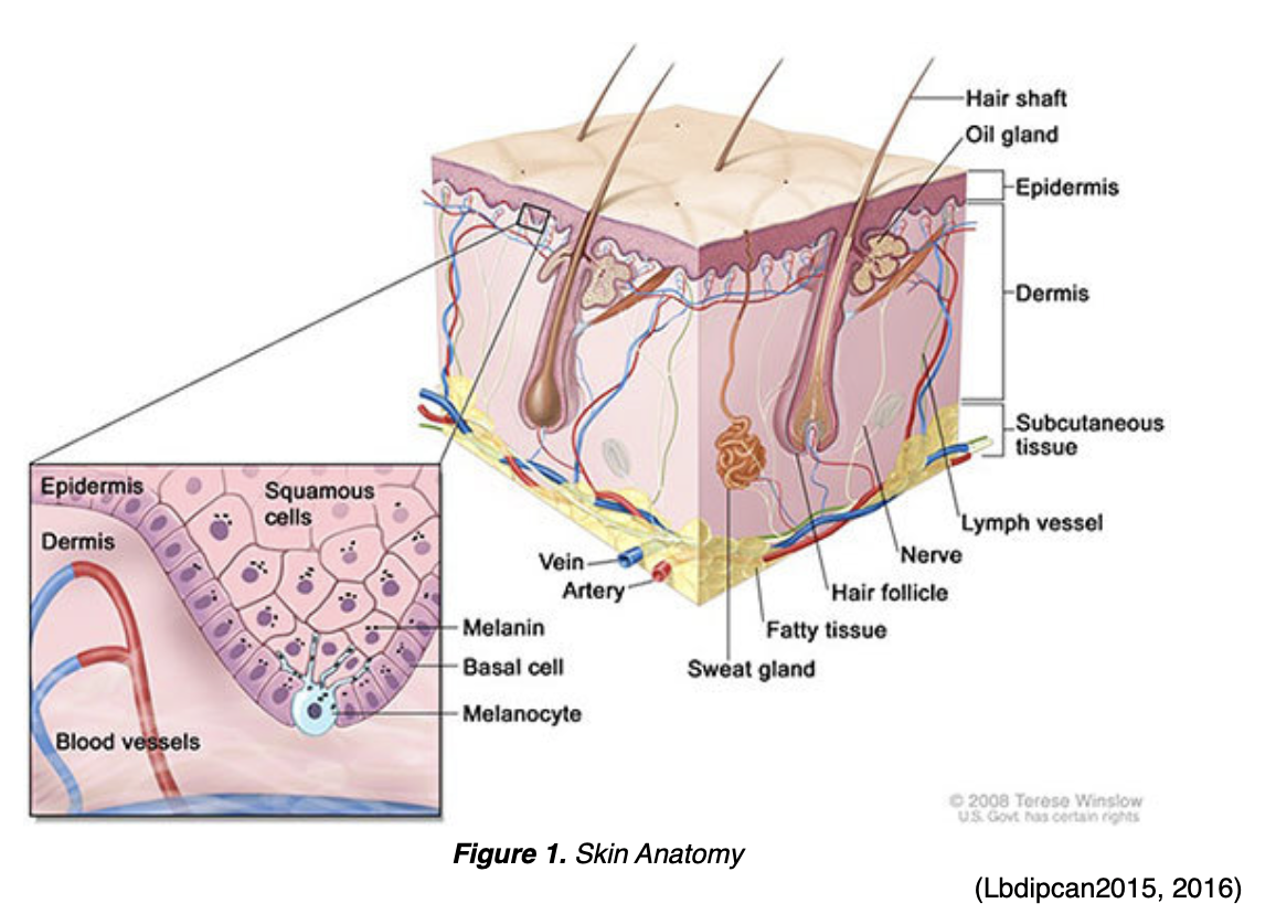
The skin is the largest organ in the human body and functions to protect against sunlight, injury, heat, and infection. It also regulates body temperature, stores water and fat, and produces vitamin D. The skin is comprised of three main layers, the epidermis, dermis, and subcutaneous tissue. Each layer of skin contains different types of cells that serve specific functions (Honari & Maibach, 2014). Figure 1 provides a graphic depiction of the layers and components of the skin.
Epidermis
The epidermis is the outermost, surface layer of the skin and functions to protect against microorganisms by providing a barrier to keep out intruders from the outside environment. The epidermis is divided into five layers as outlined below in Table 1 and is primarily made up of keratinocytes, which are flat, scale-like stratified squamous epithelial cells. Keratinocytes are responsible for the synthesis of the protein keratin, which forms a protective barrier for the body. Underlying this layer of skin are spherical-shaped basal cells, followed by the lowest, inner level of the epidermis, which contains melanocyte cells. Melanocyte cells make melanin, which is a protein pigment that is responsible for giving skin its natural color. Melanin also protects skin from the harmful effects of UV radiation damage. The more melanin present, the darker the skin color. When the skin is exposed to the sun, melanocytes produce more melanin, causing the skin to darken or tan. Clusters of melanocytes and surrounding tissue frequently create moles or nevi, which are noncancerous growths (Honari & Maibach, 2014).
Table 1. Layers of Epidermis (In order from most superficial to deepest)
Stratum Corneum | Composed of many dead skin cells that are shed into the environment; functions to repel water. |
Stratum Lucidum | Found on the palms of the hands, fingertips, and the soles of the feet. |
Stratum Granulosum | Where part of keratin production occurs. |
Stratum Spinosum | Composed of layers of keratinocytes; contains Langerhans cell which functions as a macrophage by engulfing bacteria, foreign particles, and damaged cells |
S
...purchase below to continue the course | Contains melanocytes; where keratinocytes are formed before moving up to the surface of the epidermis and being shed into the environment as dead skin cells. |
(Honari & Maibach, 2014)
Dermis
The dermis is the layer beneath the epidermis and is comprised of collagen-producing fibroblasts, which are proteins responsible for giving skin strength, durability, and flexibility. The dermis contains blood and lymph vessels, glands, hair follicles, and nerves. Blood vessels nourish the skin with oxygen and nutrients, deliver immune system cells to the skin to fight infection, and carry away waste products. Some of the glands produce sweat, which helps to regulate body temperature, whereas other glands produce sebum, an oily substance that helps keep the skin from drying out. Nerves communicate signals from the skin, including touch, pain, pressure, temperature, and itching (Honari & Maibach, 2014).
Subcutaneous Tissue
The deepest layer of the skin is the subcutaneous layer. Similar to the dermis, it contains blood vessels and nerves. It also includes a layer of fat that serves to cushion and protect against physical trauma or impact to internal organs, muscles, and bones. This layer of fat also helps regulate body temperature (Honari & Maibach, 2014).
Basics of Cancer Biology
Cancer begins in cells, which are the building blocks that make up all tissues within the human body, and tissues make up organs. In theory, any cell in the body can develop into cancer. When functioning normally, cells within the body grow and divide through a system of checks-and-balances called the cell cycle. Through this process, as cells grow old, they die and are eliminated by the body, and new cells take their place. Cancer develops when there is a disruption in the orderly process, and the cell cycle goes haywire. New cells form when the body does not need them, and old cells do not die when they should. These new cells grow uncontrollably, function abnormally, and develop into a cancerous tumor (Yarbro et al., 2018).
Risk Factors
Actinic keratosis (AK) is the most common pre-cancerous skin condition affecting more than 58 million Americans. Approximately 65% of all squamous cell carcinomas and 36% of all basal cell carcinomas arise in lesions that were previously diagnosed as actinic keratoses. AK is most commonly diagnosed in individuals who have fair skin and are older than age 40, as AK tends to develop after years of sun exposure. Approximately 90% of NMSCs are associated with exposure to UV radiation from the sun (SCF, 2019c). Among all types of skin cancer, specific risk factors associated with increased environmental or artificial UV exposure include:
- Long term, intense sun exposure lacking adequate use of sunblock and other sun safety precautions;
- Northern European ethnic origin;
- People with fair skin, blond, or red hair, and light-colored eyes;
- A tendency to burn rather than tan;
- Proximity to the equator;
- History of blistering sunburns in childhood and adolescence;
- Use of indoor tanning beds;
- People with a weakened immune system (immunocompromised state), including having had an organ transplant;
- Exposure to therapeutic ionizing radiation;
- HIV seropositive;
- Specific genetic syndromes: nevoid basal cell carcinoma and xeroderma pigmentosum (ACS, 2019b; Marzuka & Book, 2015).
Risk Factors specific to increased risk for NMSC development include:
- Prior scars, burns, ulcers, or areas of inflammation of the skin;
- Basal cell nevus syndrome (Gorlin syndrome), which is an autosomal dominant mutation of the human patched gene;
- Chronic exposure to arsenic (ACS, 2019b; SCF, 2019a, 2019c).
The sun's UV rays can damage unprotected skin in as little as fifteen minutes, yet it can take up to 12 hours for skin to show the full effect of sun exposure. Those who have already been diagnosed with skin cancer have an increased chance of developing it again, especially in the same sun-damaged area or in a nearby location. A tan is the body's attempt to protect itself from the sun's harmful rays, even without a sunburn (SCF, 2019c). Sunburns and suntans are signs that skin has been damaged. The more damage the sun does to skin, the more likely the individual is to have complications. These include early wrinkles, skin cancer, or other skin problems (Marzuka & Book, 2015).
For melanoma, major risk factors include a personal or family history of melanoma. Melanoma risk is greater if there are one or more first-degree relatives (parent, sibling, child) who have been diagnosed with melanoma. Statistically, about 10% of all people with melanoma have family members with melanoma. Another risk factor is the presence of dysplastic nevi, a type of mole that looks different from an ordinary mole. A dysplastic nevus may be bigger than a common mole, and its color, surface, and border may be different. It's usually wider than a pea and longer than a peanut. A dysplastic nevus can have a mixture of several colors, from pink to dark brown. Often, it's flat with a smooth, slightly scaly, or pebbly surface, and it has an irregular edge that may fade into the surrounding skin. A dysplastic nevus is more likely than a common mole to turn into melanoma (NCI, 2018).
An important risk factor for melanoma includes the presence of atypical, large, or numerous (more than 50) moles. Similar to NMSC lesions, melanoma is much more common in individuals of fair complexion and those who have been exposed to natural or artificial sunlight (i.e., tanning beds) (NCI, 2019). The use of artificial tanning beds before 35 years of age increases the risk for melanoma by 78% (CDC, 2017). Heavy exposure to UV radiation, from sunlight or the use of indoor tanning, is a risk factor for all types of skin cancer; indoor tanning devices are classified as carcinogenic by the International Agency for Research on Cancer. One blistering sunburn during childhood doubles the risk of melanoma, whereas five or more blistering sunburns before age 20 increases the risk of melanoma by 80% (ACS, 2019b).
Ultraviolet Radiation
UV radiation is part of the natural energy produced by the sun's invisible rays but is also produced from artificial sun lamps and tanning beds. UV radiation can damage skin and lead to all types of skin cancer. On the electromagnetic spectrum, UV light has shorter wavelengths than visible light, so it cannot be seen with the naked eye, but the skin can feel it and be impacted by it. Two types of harmful UV radiation reach the Earth's surface and are proven to contribute to the risk of skin cancer; long-wave ultraviolet A (UVA) and short-wave ultraviolet B (UVB) rays. While UVA and UVB rays differ in how they affect the skin, they both induce harm to the skin, and when combined, the risk for damage rises. Unprotected exposure to both UVA and UVB rays damages the DNA in skin cells, producing genetic defects, or mutations, that can lead to premature aging, skin cancer, and suppression of the immune system. The more prolonged and more intense the exposure, the higher the risk of harmful consequences to the skin (Itano, 2016). Table 2 provides an outline of the differences between these two types of rays.
Table 2. UVA versus UVB Radiation Rays
UVA Radiation | UVB Radiation |
Has a longer wavelength and is associated with skin aging. | Has a shorter wavelength and is associated with skin burning. |
Damages the collagen and elastin in the skin, generates free radicals, and causes almost all forms of skin aging, including wrinkles (photoaging). | Plays a key role in the development of skin cancer by damaging cells and causing DNA mutations; can eventually lead to melanoma and cause cataracts. |
Able to penetrate deep into the dermis of the skin, resulting in a tan, and can penetrate through glass. | Does not penetrate as deep as UVA but is the primary source of sunburn. Usually burns the superficial layers of the skin. |
Makes up 95% of all the UV rays that make it to the Earth’s surface. | UVB radiation makes up only 5% of the UV rays from the sun, but it is very high energy. |
Primary source of radiation used in tanning beds. UV rays produced by tanning units are 10–15 times more powerful than UV rays produced by midday sun. | Intensity of UVB rays vary by season, geographic location, and time of day, with 10 AM to 4 PM considered the peak hours. |
(Saric-Bosanac et al., 2019).
UV radiation can penetrate light clothing, windows, and windshields. It is also reflected by sand, water, snow, and concrete, and can reach swimmers at least 1 foot below the surface of the water. Roughly 60% to 80% of the sun's rays go through clouds, so clouds and water do not offer protection (Saric-Bosanac et al., 2019).
Screening and Early Detection
The best way to detect skin cancer early is to be aware of new or changing skin growths, particularly those that look unusual. The ACS (2019a) recommends that any new lesion or a progressive change in a lesion's appearance (size, shape, or color change) should be evaluated promptly by a clinician. While many researchers and clinicians argue that skin cancer screening with a total body skin examination (TBSE) is a safe and cost-effective screening test, there remains no national consensus regarding its benefit or implementation. The USPSTF (2016) recommendation statement concludes that the current evidence is "insufficient to assess the balance of benefits and harms of visual skin examination by a clinician to screen for skin cancer in adults" (p. 479). This recommendation statement only applies to asymptomatic adults who do not have a history of pre-cancerous or cancerous skin lesions. Therefore, total body skin examination is not considered part of the general physical examination performed by primary care providers or other non-dermatology specialties (USPSTF, 2016).
Skin Self-Examination
While the USPSTF (2018) concludes that the current evidence is insufficient regarding self-examination to prevent skin cancer, many organizations including the American Academy of Dermatology (AAD, 2019), the ACS (2019c), and the Melanoma Research Alliance (n.d.), jointly emphasize the importance of monthly skin self-examinations for the detection of early skin cancer. The purpose of teaching routine self-examination of the skin is to promote the early detection of skin cancer. Engaging in self-care activities contributes to the maintenance of health. These organizations encourage that patients are counseled to perform skin self-examination routinely, preferably every month in a well-lit room during daylight hours. Patients will need a wall mirror and a hand mirror, as well as a chair footrest to perform an adequate self-assessment. Patients should also be given access to a body map diagram or encourage the use of a skin mole tracking mobile device application to note any areas of suspicion. Patients should be counseled to bring the body map diagram to their clinician for evaluation of any possible suspicious lesions (AAD, 2019). Figure 2 outlines the proper steps to take when performing a skin cancer self-examination. The AAD (n.d.a) started a campaign called SPOT Skin Cancer, offering a free body map tool for patients to use, as displayed in Figure 3.
Figure 2. How to Perform a Skin Self-Exam 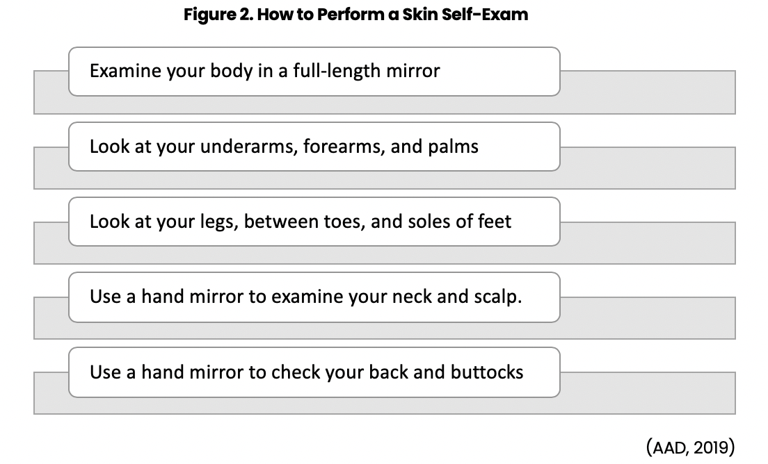
Figure 3. DETECT Skin Cancer: Body Mole Map

Signs and Symptoms
Generalized Warning Signs
Warning signs of all skin cancers include changes in the size, shape, or color of a mole or other skin lesion, the appearance of new growth on the skin, or a sore that doesn't heal. Changes that progress over a month or more should always warrant evaluation by a health care provider(ACS, 2019b).
The ABCDE Rule
Dermatologists routinely perform TBSE, assessing skin lesions utilizing the ABCDE rule. The ABCDE rule outlines specific characteristics to be aware of when assessing suspicious abnormalities. This rule provides an easy way to recognize moles and growths that may be cancerous. Melanoma can disguise itself in the form of a suspicious mole or lesion, so understanding the ABCDE rule can save lives. However, not all melanomas have all of these signs, so it is important to be alert for any new or changing skin growths or spots (ACS, 2019b; Itano, 2016). Table 3 provides a summary and overview of the clinical use of the ABCDE rule, and Figure 4 demonstrates melanoma lesions compared to normal moles, or benign nevi.
Table 3. ABCDE Rule of Melanoma
A Asymmetry | A is for Asymmetry
|
B Border Irregularity | B is for Border Irregularity
|
C Color | C is for Color
|
D Diameter | D is for Diameter
|
E Evolution | E is for Evolution
(ACS, 2019b) |

Actinic Keratosis
Actinic Keratosis most commonly appears on sun-exposed areas of the skin, such as the head, neck, forearms, and hands. They develop as dry, scaly patches or spots. Although this skin condition is pre-cancerous, it is a strong precursor to the development of SCC and BCC. Therefore, early identification and treatment remain important (ACS, 2019b).
Basal Cell Carcinoma
Basal cell carcinomas arise from abnormal, uncontrolled growth of basal cells within the epidermis. BCC lesions tend to be slow-growing and are rarely fatal, as it is incredibly uncommon for them to spread to other parts of the body. However, these lesions can be highly disfiguring if allowed to grow and can cause extensive invasion of the surrounding tissues. BCC can grow into the nerves and bones, causing damage and disfigurement. BCC lesions often develop on regions of the body that receive regular sun exposure such as the face and hands but can form anywhere on the body, including the chest, abdomen, and legs. They generally appear as flesh-colored, pearly bumps or a raised pink or red translucent, shiny area. On dark skin, BCC can present in tan, black, or brown colors and can easily be mistaken for a normal mole. Alternatively, they may present as an open sore, scar, or growth with rolled edges or a central indentation. These lesions may develop tiny surface blood vessels over time. Lesions may crust, ooze, itch, and often bleed following minor injury. To avoid misdiagnosing a lesion, healthcare professionals must understand that BCC lesions may appear quite different from one individual to another (SCF, 2019a).
According to the Skin Cancer Foundation (2019b), the five most common warning signs of BCC include:
- An open sore that does not heal, or may appear to heal, but then reappears;
- A reddish patch or irritated area;
- A shiny bump or nodule;
- A small pink growth;
- A scar-like area that is flat white, yellow, or waxy in color. The skin may be shiny and taut with poorly defined borders, indicating invasive BCC (SCF, 2019a).
There are four common subtypes of BCCs, which are characterized by distinctive features, as outlined below in Table 4.
Table 4. Four Common Types of BCC and their Associated Characteristics
Nodular basal cell carcinoma | Pigmented basal cell carcinoma | Morpheaform or sclerotic basal cell carcinoma | Superficial basal cell carcinoma |
|
|
|
|
(Itano, 2016).
Squamous Cell Carcinoma
Squamous cell carcinomas arise from the keratinocyte cells within the epidermis, and while these lesions are less common than BCC, they are more likely to spread. SCC are round to irregular shapes with a plaque-like or nodular character. The appearance of SCC can vary from an ulcerated, infiltrating mass to an elevated erythematous nodular mass. Additionally, SCC lesions may feel rough, scaly or lumpy, and blood vessels may appear at the edge, causing them to bleed easily. SCC most commonly appears on parts of the body that experience increased levels of sun exposure such as the face, lips, and back (Itano, 2016).
Bowen disease is an intraepithelial form of SCC and is also referred to as squamous cell carcinoma in situ. Bowen disease is associated with HPV and is confined to the epidermis. It does not infiltrate the dermis and often presents as a rough, scaly, erythematous plaque with irregular borders. Most cases present on the lower legs and sun-exposed skin and are primarily found in women (Linares et al., 2015).
Melanoma
Melanoma arises from the melanocyte cells and is characterized by radial and vertical growth. While the majority of melanoma is referred to as cutaneous melanoma, appearing in the skin, melanoma may also occur in the eye, known as ocular melanoma. There are rare instances where melanoma arises in other areas such as the lymph nodes or digestive tract. Melanoma can develop on any skin surface but is most commonly found on the trunk, head, and neck in males and on the lower legs and feet in females. The classification of cutaneous melanoma lesions is outlined below in Table 5 and includes four major subtypes: superficial spreading melanoma, nodular melanoma, lentigo maligna melanoma, and acral-lentiginous melanoma (Itano, 2016).
Table 5. Classification of Malignant Melanoma
Superficial spreading melanoma (SSM) | Nodular melanoma (NM) | Lentigo maligna melanoma (LMM) | Acral-lentiginous melanoma (ALM) | |
Common sites |
|
|
|
|
Features |
|
|
|
|
(Itano, 2016).
Melanoma is rare in those who have naturally dark-skinned complexion, such as African Americans, although when it does occur, it is usually beneath the fingernails or toenails, or on the palms or soles. Ultimately, melanoma can metastasize throughout the whole body, but most commonly spreads to the liver, lungs, and brain. Upon arrival at these locations, the cancerous cells replicate and develop into malignant tumors and can ultimately induce death if left untreated (Itano, 2016).
Box 1-1. Critical Consideration. 
Diagnosis
Early diagnosis and treatment for skin cancer are vital, as nearly all NMSC lesions can be successfully treated through excision without complication. Early identification and treatment of melanoma can significantly reduce the risk of spread to other tissues through the lymphatic system and blood vessels. As the most important risk factor for skin cancer is chronic UV exposure, it is vital to obtain a detailed history from the patient. Diagnosis is usually suspected in older, fair-skinned patients who present with scaly, indurated lesions on sun-exposed areas of the head and neck. The accuracy of clinical diagnosis can be enhanced using adequate room lighting and a dermatoscope. A dermatoscope is a handheld tool that offers an inbuilt illuminating system and magnifying optics to facilitate the visualization of subsurface skin structures that are not easily detectable to the unaided eye. The application of an immersion fluid such as mineral oil, ultrasound gel, or liquid paraffin to the skin can enhance translucency, improving the visibility of the subsurface skin structures of the lesion under investigation (Sonthali & Kaliyadan, 2019).
In addition to information acquired from a thorough history and physical examination, most early skin cancers are diagnosed and treated by removal (excision), followed by microscopic examination of the cells. Histopathology is the gold standard for definitive diagnosis of skin cancer. A biopsy should be performed for any lesion that is suspicious of melanoma. Several types of skin biopsies may be performed, such as a punch biopsy, shave biopsy, and excisional biopsy. For the diagnosis of melanoma, a full-thickness biopsy is required because management and prognosis depend upon the depth of the lesion (Linares et al., 2015).
Punch Biopsy
A punch biopsy is performed to excise small lesions ranging from 2 mm to 10 mm in size. It is not a sterile procedure and is usually performed in the office setting. The site is prepared with isopropyl alcohol, povidone-iodine, or chlorhexidine and anesthetized with 1% to 2% lidocaine. A disposable round knife punch tool is often used, and the biopsy is generally taken to the depth of the subcutaneous tissue. For the larger punch biopsies (those 8 to 10 mm), one or two sutures may be used for cosmetic results. Patients should be advised to keep the area clean and dry, and to apply an antibiotic ointment (such as bacitracin [Neosporin]) twice daily (Linares et al., 2015).
Shave Biopsy
A shave biopsy is also performed in the office and is not a sterile procedure. The site is prepped in the same manner as for the punch biopsy, but a wheal of anesthesia may be injected to raise the lesion to assist in the biopsy. The shave biopsy is performed with a 15-blade scalpel or double-edged razor tool. It is the recommended choice of biopsy for lesions that are confined to the epidermis. It should not be performed for pigmented lesions or those that are suspicious for melanoma because the depth of the lesion is vital for melanoma staging purposes. Sutures are not necessary, and the patient should be counseled on keeping the area clean and dry and applying bacitracin (Neosporin) twice daily (Linares et al., 2015).
Excisional Biopsy
An excisional biopsy is a sterile procedure that is ideal for lesions that are large, known to be malignant, or are suspicious of malignant melanoma. An excisional biopsy includes removal of the entire lesion with 1-mm to 3-mm margins of healthy skin (called negative margins), and part of the subcutaneous fat whenever possible. Depending on the size and extent of the lesion in question, this procedure may be performed in an outpatient surgical center, operating room, or a dedicated surgical office setting with the proper equipment to ensure sterility of the procedure. Excisional biopsies are usually performed using a scalpel, and the biopsy is taken to at least the depth of the subcutaneous tissue or the extent of the visible lesion. Sutures are usually necessary, and postoperative care generally includes keeping the wound clean, dry, and applying bacitracin (Neosporin) with dressing changes (Linares et al., 2015).
Nursing Considerations
Contraindications to skin biopsies may include infection at the site, history of a bleeding disorder, or current/very recent use of blood thinners. Patients should not be taken off therapeutic or prophylactic blood thinners for a skin biopsy. Patients presenting with suspicious lesions on the face, eyelids, or lips may need to be referred to a dermatologist or plastic surgeon due to the cosmetic aspect of removing one of these lesions (Linares et al., 2015).
NMSC Treatment
Treatment for skin cancer depends on several factors, such as the type of cancer, location, identifying features, depth of invasion (if it has spread to other areas), and so forth. Most cases of NMSC are cured by removing the lesion through surgical excision, laser therapy, or deep cryosurgery (e.g., destruction of the cancerous lesion by freezing). Radiation therapy and certain topical medications may also be used. Since Bowen disease is confined to the epidermis, unlike invasive SCC, effective treatment can be seen with cryotherapy, cauterization, and topical 5-fluorouracil (Efudex, Carac). It is important to note that NMSCs can recur, even after complete surgical removal. Some cancer cells may remain undetectable after surgery, and others can form roots that extend beyond what's visible. BCCs on the nose, ears, and lips are more likely to recur, usually within the first two years after surgery. Therefore, appropriate follow-up and surveillance are crucial (Linares et al., 2015).
Curettage and electrodesiccation is a treatment option performed for the management of superficial NMSCs. In this treatment, the cancerous lesion is removed by scraping it with a curette, which is a long, thin instrument with a sharp looped edge on one end. The area is then treated with an electrode to destroy any remaining cancer cells. Mohs surgery is considered the most effective technique for treating many BCC and SCC lesions, with up to 99% cure rate for a skin cancer lesion that has not been treated before, and up to 94% for skin cancer that has recurred after prior treatment. Mohs surgery is performed in stages, but all in one visit. The surgeon removes a layer of tissue then examines it under the microscope. If any cancer cells remain, the surgeon knows the precise location where they are and removes another layer of tissue, while sparing as much healthy tissue as possible. This process is repeated until no cancer cells remain. It allows the removal of all cancerous cells for the highest cure rate while sparing healthy tissue and leaving the smallest possible scar (SCF, 2019b).
Melanoma Staging and Treatment
Once a diagnosis of melanoma has been made by biopsy of the lesion, pathologic staging must be completed to determine prognosis and treatment. The American Joint Committee on Cancer (AJCC) staging system is used to stage melanoma skin cancer, and the National Comprehensive Cancer Network (NCCN, 2019) offers evidence-based treatment guidelines correlating with each stage. In this system, there are five stages of melanoma, which are outlined below in Table 6.
Table 6. The Five Stages of Melanoma
Stage 0 |
|
Stage I |
|
Stage II |
|
Stage III |
|
Stage IV |
|
(Melanoma Research Alliance, n.d.; NCCN, 2019).
Treatment of Advanced and Metastatic Melanoma
As outlined above in Table 6, surgical intervention with wide local excision is often curative if the primary growth and surrounding healthy tissue are removed (negative margins) for localized melanoma. These patients require close follow-up and surveillance with TBSE by a skilled clinician every three months and are counseled to perform monthly skin self-examinations at home. They generally do not require additional treatment. For some patients, a sentinel lymph node is biopsied to determine the stage of the disease, and more extensive lymph node surgery may be necessary if the sentinel lymph node contains cancer. Treatment for advanced disease and metastatic melanoma can be more complex. Melanomas with deep invasion or those that have spread to lymph nodes may be treated with surgery, immunotherapy, and/or radiation therapy. The treatment for advanced melanoma has changed dramatically in recent years with FDA approval of several new immunotherapy and targeted drugs. Chemotherapy may be used at times but is generally considered much less effective than newer immune-based treatments and targeted therapies (ACS, 2019b; NCCN, 2019).
Treatment options beyond surgery may include one or more of the following modalities:
- Radiation Therapy: high-energy rays like x-rays shrink or kill the cancerous cells;
- Chemotherapy: systemic treatment administered in pill form or injected into the veins and devised to shrink or kill the cancer;
- Immunotherapy and vaccine therapy: systemic treatment injected into the body that uses substances produced by living organisms made in the body or in the lab. These are used to boost the immune system to help the body fight the cancer through the power of the immune system;
- Targeted therapy: systemic treatment administered in pill form or injected into the veins that are designed to target specific mutations or weak areas in the cancer cells;
- Clinical trial participation (Melanoma Research Alliance, n.d.; NCCN, 2019).
Skin Cancer Follow-up Care
Patients who have been diagnosed with NMSC or melanoma skin cancer and completed treatment still require follow-up surveillance at regular intervals. For patients who had BCC, follow-up is often recommended every 6 to 12 months. For those who had SCC, monitoring is usually more frequent than with BCC, often every three to six months for the first few years. If the patient remains without any evidence of cancerous lesions, their follow-up intervals are extended to a longer time between each visit (ACS, 2019d). Surveillance for melanoma is much more complicated. It depends on the stage of cancer at diagnosis, response to treatment, the extent of residual disease (if any), and any need for continued treatment. Melanoma-in-situ treated with wide local excision with negative margins is considered curative treatment. The NCCN advises these patients to follow up annually. For patients with stage IIB-IV who are without evidence of disease, or in remission, the NCCN recommends follow up every three to six months for two years, then every three to twelve months for three years, and then annually as clinically indicated. For all patients with a prior history of melanoma, the NCCN guidelines recommend at least annual skin exams for life (NCCN, 2019).
Skin Cancer Prevention and Sun Safety
Minimizing sun exposure is the key to preventing skin cancer. According to American Cancer Society researchers, most melanoma cases and deaths are potentially preventable by minimizing exposure to intense and damaging UV radiation. The USPSTF (2018) found moderate grade evidence to recommend counseling young adults, adolescents, children, and patients of young children to minimize exposure to UV radiation for persons aged six months through 24 years with fair skin to reduce their risk of skin cancer. Further, the USPSTF recommends clinicians selectively offer counseling to adults older than 24 years with fair skin about minimizing their exposure to UV radiation. Existing evidence indicates that the net benefit of counseling all adults older than 24 years is small. In determining whether this service is appropriate in individual cases, patients and clinicians should consider the presence of other risk factors for skin cancer (USPSTF, 2018). Nevertheless, below is a compilation of evidence-based interventions to educate patients regarding reducing the risk of skin cancer induced by UV radiation.
Sun-Safety Guidelines to Share with Patients
- Avoid sunbathing or indoor tanning;
- Seek shade when outdoors; however, understand that while the shade is an effective mode of reducing UV radiation, it does not eliminate the exposure completely;
- Avoid exposures to midday sun when the sun's UV rays are strongest and can do the most damage to the skin (from 10 AM through 4 PM);
- When outside, wear protective clothing made of tightly-woven fabric (e.g., long sleeves, a wide-brimmed hat, long pants, etc.). A hat with at least a 6-inch brim around the entire circumference is recommended as it offers protection to the face, neck, and ears. Baseball caps do not protect the back of the neck or the tops of the ears;
- Wear sunglasses with UV-absorbing lenses to block UV rays; seek sunglasses that protect the sides of eyes and lenses that offer protection against both UVA and UVB rays. Sun exposure increases the risk of cataracts;
- Apply broad-spectrum sunscreen that has a sun protection factor (SPF) of at least 30 to unprotected skin every day, regardless of anticipated sun exposure and even on cloudy days;
- Adequate sun protection should be ensured while driving or sitting near windows due to exposure to UVA and UVB rays through the glass; especially to the hands, arms, neck, and face;
- Be mindful of medications, as there are some over-the-counter and prescription medications that can increase photosensitivity (sensitivity to harmful sun rays). A few of the most common include:
- Antihistamines such as diphenhydramine (Benadryl),
- Ibuprofen (Motrin),
- Certain antibiotics such as tetracyclines (doxycycline [Monodox] and minocycline [Minocin]),
- Certain antidepressant medications such as selective serotonin reuptake inhibitors (fluoxetine [Prozac] or citalopram [Celexa]),
- Several types of chemotherapeutic agents, such as capecitabine (Xeloda), doxorubicin (Adriamycin), or fluorouracil (5-FU) (SCF, 2019c).
Understanding SPF
The AAD (n.d.b) recommends that everyone use broad-spectrum sunscreen, which provides protection against UVA and UVB rays. Sunscreens are rated in strength, according to SPF. The higher the SPF, the more sunburn protection provided. Sunscreen should be SPF 30 or higher and water-resistant. The AAD cautions that sunscreen does not provide 100% protection against the sun's harmful UV rays, so the above precautions must still be taken to ensure safety.
Sunscreen Tips
- Use sunscreen every day, even on cloudy days. Up to 80% of the sun’s harmful UV rays can penetrate skin through the clouds;
- Snow, sand, and water increase the need for sunscreen as they reflect the sun’s rays;
- Apply enough sunscreen to cover all exposed skin. Most adults need about 1 ounce to fully cover their body;
- Rub sunscreen in well to dry skin;
- Reapply at least every two hours when outside, and more frequently after swimming, sweating, or toweling off;
- Ensure the sunscreen has not expired, as some ingredients in sunscreen break down over time;
- Sunscreen should be applied at least 15 minutes prior to sun exposure to all areas that the sun’s rays may reach. The ACS recommends special attention to the areas most commonly omitted when applying sunscreen, including the tips of ears, the back of neck, and bald spots on the top of the head;
- Apply protective lip treatment with SPF of at least 15 to protect the lips from sun exposure;
- Type of sunscreen
- Products that include zinc oxide generally offer the safest sun protection;
- However, the AAD advises that the best type of sunscreen is the one the patient you will use over and over. It should be broad-spectrum (UVA and UVB), with an SPF of 30 or higher, and water-resistant;
- Available sunscreen options include lotions, creams, gels, ointments, wax sticks and sprays;
- Creams are best for dry skin and the face;
- Gels are ideal for hairy areas, such as the scalp or male chest;
- Sticks are good to use around the eyes;
- Sprays are sometimes preferred by patients since they are easy to apply. However, current FDA regulations on testing and standardization do not pertain to spray sunscreens, so the AAD advises against their use (AAD, n.d.b).
Vitamin D
Vitamin D is an essential nutrient that is needed for healthy bones, muscles, and teeth. The human body makes vitamin D whenever it is exposed to sunlight (specifically UVB, which is processed through the skin). While sunlight is important for vitamin D, patients must be counseled on the importance of being careful about how much sunlight they are getting. In general, the lighter the skin tone, the less sunlight needed on the skin to make vitamin D. The body only needs about 15 minutes of unimpeded sun exposure, two to three times a week, to fulfill the body's vitamin D needs. This small amount of sun exposure generates all the vitamin D the body requires. After that, the body starts to dispose of vitamin D to avoid an overload of the vitamin, at which point sun exposure is providing patients with risk yet without any of the presumed benefits. In addition to sunlight, vitamin D can be acquired through one's diet. Diets that include egg yolks, oily fish (i.e., salmon, tuna, sardines, eel), or fortified milk and cereal can satisfy vitamin D needs without sun exposure. Alternatively, patients can be advised to take vitamin D supplements (SCF, 2016).
Special Considerations for Children and Adolescents
As described earlier, sunburns in childhood significantly increase the risk of melanoma later in life. Children who are most vulnerable to harmful UV effects are those with fair skin, red or blonde hair, freckles, and light-colored eyes. Use of certain medicines (such as antibiotics) and tanning beds increase a child's or adolescent's risk for harmful effects of the sun. Sun avoidance/limiting exposure to sunlight and sun protection measures can be used to reduce the risk of harmful effects of overexposure. Parents should be taught to generously apply sunscreen with an SPF of at least 30 to their child's body, cover exposed skin with about 1 ounce of sunscreen 15 to 20 minutes prior to going outside, and reapply every two hours, even on cloudy days, and after swimming or sweating. Infants younger than six months of age should never be outside in direct sunlight and should be dressed in tightly woven long-sleeved shirts, long pants, and brimmed hats which offer the best protection. Traditionally, it was advised to avoid applying sunblock to infants under six months, but the American Academy of Pediatrics (AAP) has now published new guidelines, as research has shown that the risk of sunburn is greater than the risk of chemical exposure. When adequate clothing and shade are not available, parents are advised to apply a minimal amount of sunscreen with at least 15 SPF on infants under six months (AAP, 2019). Clothing, especially swimsuits, can be manufactured with SPF fabric. There are also small tents and umbrellas that are designed with SPF 50 fabric to provide protection to the entire family simultaneously when seated underneath it with no chemical exposure at all.
References
American Academy of Dermatology. (n.d.a). Infographic: Skin cancer body mole map. Retrieved October 11, 2019 from https://www.aad.org/diseases/skin-cancer/body-mole-map
American Academy of Dermatology. (n.d.b). Sunscreen FAQS. Retrieved October 10, 2019 from https://www.aad.org/sun-protection/sunscreen-faqs
American Academy of Dermatology. (2019). Detects skin cancer: How to check your skin. https://www.aad.org/skin-cancer-find-check
American Academy of Pediatrics. (2019). Sun safety and
protection tips from the American Academy of Pediatrics. https://www.aap.org/en-us/about-the-aap/aap-press-room/news-features-and-safety-tips/Pages/Sun-Safety-and-Protection.aspx
American Cancer Society. (2019a). About basal and squamous cell skin cancer. http://www.cancer.org/cancer/basal-and-squamous-cell-skin-cancer/about.html
American Cancer Society. (2019b). Cancer facts & figures. https://www.cancer.org/content/dam/cancer-org/research/cancer-facts-and-statistics/annual-cancer-facts-and-figures/2019/cancer-facts-and-figures-2019.pdf
American Cancer Society. (2019c). How to do a skin self-exam. https://www.cancer.org/healthy/be-safe-in-sun/skin-exams.html
American Cancer Society. (2019d). Living as a basal or squamous cell skin cancer survivor. https://www.cancer.org/cancer/basal-and-squamous-cell-skin-cancer/after-treatment/follow-up.html
Centers for Disease Control and Prevention. (2017). How can I protect my children from the sun? https://www.cdc.gov/cancer/skin/basic_info/children.htm
Centers for Disease Control and Prevention. (2019). Skin cancer. https://www.cdc.gov/cancer/skin/index.htm
Cleveland Clinic. (2015). Skin cancer: Why you should be concerned. https://my.clevelandclinic.org/health/articles/8315-skin-cancer-why-you-should-be-concerned
Honari, G., & Maibach, H. (2014). Chapter 1 – Skin structure and function. In Applied dermatotoxicology [E-reader version] (pp. 1-10). https://www.sciencedirect.com/science/article/pii/B9780124201309000013
Itano, J. K. (2016). Core curriculum for oncology nursing. (5th ed.). (J. Brant, F. Conde, & M. Saria, Eds.). Elsevier.
Linares, M. A., Zakaria, A., & Nizran, P. (2015). Skin cancer. Primary Care Clin Office Pract, 42, (4), 645-659. https://doi.org/10.1016/j.pop.2015.07.006
Marzuka, A. G., & Book, S. E. (2015). Basal cell carcinoma: Pathogenesis, epidemiology, clinical features, diagnosis, histopathology, and management. Yale Journal of Biology & Medicine, 88(2), 167-179.
Melanoma Research Alliance. (n.d.). Melanoma staging. Retrieved October 12, 2019 from https://www.curemelanoma.org/about-melanoma/melanoma-staging/
National Comprehensive Cancer Network. (2019). Cutaneous melanoma. https://www.nccn.org/professionals/physician_gls/pdf/cutaneous_melanoma.pdf
National Cancer Institute. (2018). Common moles, dysplastic nevi, and risk for melanoma. https://www.cancer.gov/types/skin/moles-fact-sheet
National Cancer Institute. (2019). SEER cancer statistics review, 1975-2016. https://seer.cancer.gov/csr/1975_2016/results_merged/sect_16_melanoma_skin.pdf
Saric-Bosanac, S. S., Clark, A. K., Nguyen, V., Pan, A., Chang, F., Li, C., & Sivamani, R. K. (2019). Quantification of ultraviolet (UV) radiation in the shade and in direct sunlight. Dermatology Online Journal, 25(7), 1-6.
Siegel, R.L., Miller, K.D., & Jemal, A. (2019). Cancer statistics, 2019. CA Cancer Journal for Clinicians, 69(1), 7-34. https://doi.org/10.3322/caac.21551.
Skin Cancer Foundation. (2016). Sun protection and vitamin D. https://www.skincancer.org/blog/sun-protection-and-vitamin-d/
Skin Cancer Foundation. (2019a). Basal cell carcinoma warning signs. https://www.skincancer.org/skin-cancer-Information/basal-cell-carcinoma/bcc-warning-signs-images/
Skin Cancer Foundation. (2019b). Mohs surgery. https://www.skincancer.org/treatment-resources/mohs-surgery/
Skin Cancer Foundation. (2019c). Skin cancer facts & statistics. https://www.skincancer.org/skin-cancer-information/skin-cancer-facts/
Sonthali, S., & Kaliyadan, F. (2019). Dermoscopy overview and extradiagnostic applications. StatPearls [Internet]. National Institutes of Health. https://www.ncbi.nlm.nih.gov/books/NBK537131/
Stevenfruitsmaak. (2008). Melanoma lesions (left) compared to benign nevi (right) [image]. https://upload.wikimedia.org/wikipedia/commons/4/43/Melanoma_vs_normal_mole_ABCD_rule_NCI_Visuals_Online.jpg
U.S. Preventative Services Task Force. (2016). Screening for skin cancer: US preventive services task force recommendation statement. JAMA, 316(4), 429-435. https://doi.org/10.1001/jama.2016.8465
U.S. Preventative Services Task Force. (2018). Behavioral counseling to prevent skin cancer US preventative services task force recommendation statement. JAMA, 319(11), 1134-1142. https://doi.org/10/1001/jama.2018.1623
Yarbro, C. H., Wujcik, D., & Gobel, B. H. (Eds.). (2018). Cancer nursing: Principles and practice. (8th ed.). Jones & Bartlett Learning.